. 2008 Jun;135(12):2193-202.
doi: 10.1242/dev.016378. Epub 2008 May 14.

Cell-autonomous requirement for beta1 integrin in endothelial cell adhesion, migration and survival during angiogenesis in mice

Affiliations

Cell-autonomous requirement for beta1 integrin in endothelial cell adhesion, migration and survival during angiogenesis in mice

Timothy R Carlson et al. Development. 2008 Jun.

Abstract

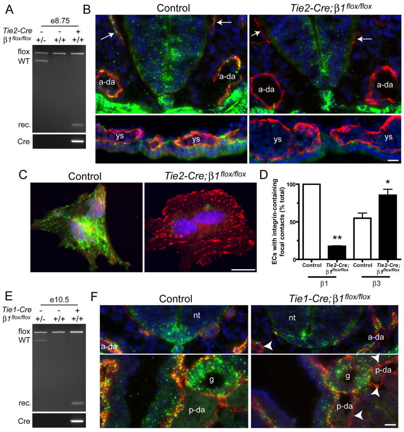
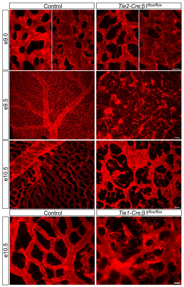
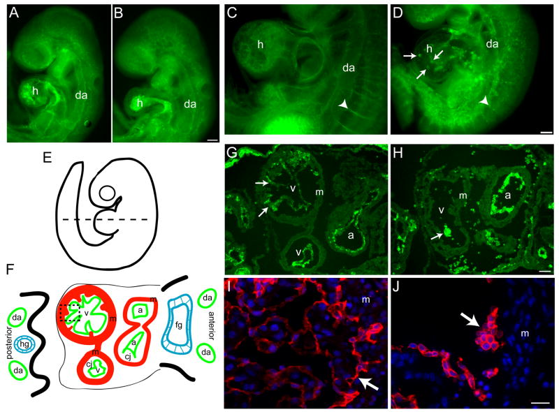
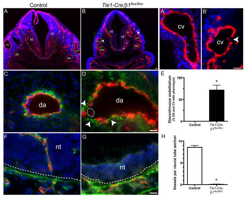
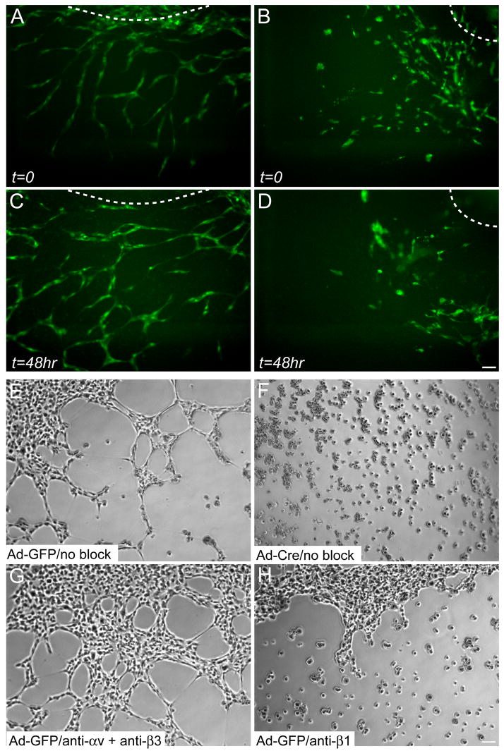
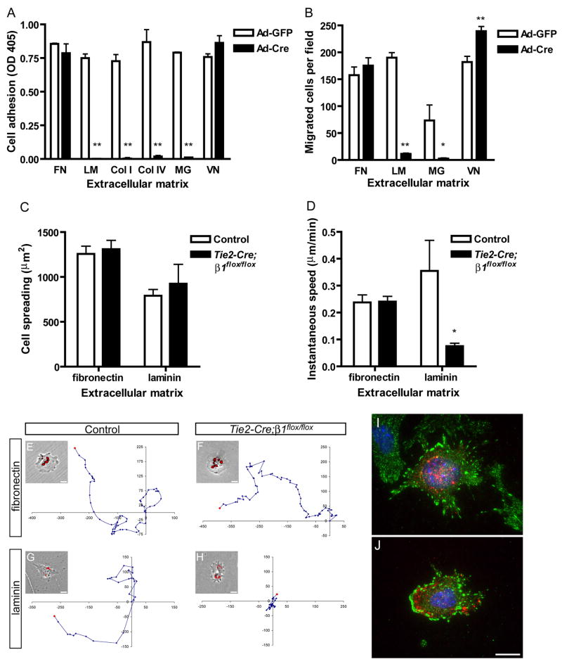
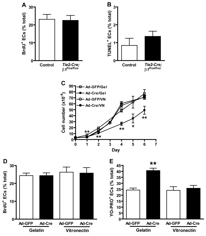
beta1 integrin (encoded by Itgb1) is established as a regulator of angiogenesis based upon the phenotypes of complete knockouts of beta1 heterodimer partners or ligands and upon antibody inhibition studies in mice. Its direct function in endothelial cells (ECs) in vivo has not been determined because Itgb1(-/-) embryos die before vascular development. Excision of Itgb1 from ECs and a subset of hematopoietic cells, using Tie2-Cre, resulted in abnormal vascular development by embryonic day (e) 8.5 and lethality by e10.5. Tie1-Cre mediated a more restricted excision of Itgb1 from ECs and hematopoietic cells and resulted in embryonic lethal vascular defects by e11.5. Capillaries of the yolk sacs were disorganized, and the endothelium of major blood vessels and of the heart was frequently discontinuous in mutant embryos. We also found similar vascular morphogenesis defects characterized by EC disorganization in embryonic explants and isolated ECs. Itgb1-null ECs were deficient in adhesion and migration in a ligand-specific fashion, with impaired responses to laminin and collagens, but not to fibronectin. Deletion of Itgb1 reduced EC survival, but did not affect proliferation. Our findings demonstrate that beta1 integrin is essential for EC adhesion, migration and survival during angiogenesis, and further validate that therapies targeting beta1 integrins may effectively impair neovascularization.


Figures

Figure 1
Figure 1. Tie2- and Tie1-Cre mediate efficient deletion of β1 integrins in embryos
Gene deletion analyses of Tie2- (AD) and Tie1- (E,F) mutants. (A) Genomic PCR analysis of e8.5 embryos demonstrates recombination (rec.) of β1 in embryos carrying Tie2-Cre and two floxed alleles of β1. (B) e8.5 cryosections were stained with anti-CD31 (red), anti-β1 integrin (Ha2/5, green), and DAPI (blue). (C) Collagenase-dissociated e8.5 embryonic cells were plated onto FN and stained with anti-β1 (HMβ1-1, green), anti-β3 (red), and DAPI (blue). EC identity in C was determined by co-staining with anti-CD31 (not shown). (D) Focal contacts in isolated ECs. Bars are means + SEM of 2 (β1) or 3 (β3) experiments. **p < 0.01 by one sample T-Test and *p < 0.05 by Student’s T-test. (E) Genomic PCR analysis of e10.5 embryos from Tie1-Cre matings. (F) e9.5 cryosections were stained with anti-CD31 (red), anti-β1 integrin (green), and DAPI (blue). Arrows, primary head veins; arrowheads, β1-negative endothelium; ys, yolk sac blood islands; a-da, anterior dorsal aortae; p-da, posterior dorsal aortae; nt, neural tube; g, gut; ve, visceral endoderm. Also see Fig. 3F for diagram of embryonic structures. Bars, 20 μm.
Figure 2
Figure 2. Endothelial deletion of β1 integrins causes EC disorganization leading to cardiovascular defects and lethality at midgestation
Whole mount anti-CD31 immunostained yolk sacs at the indicated embryonic stages. The left panels of e9.0 are capillary regions and the right panels are regions apparently undergoing arteriovenous remodeling. Note the disconnected capillaries and overall vascular disorganization in all mutants. Images are representative of at least 5 control/mutant embryo pairs at each stage. Bars, 100 μm (e9.5), 50 μm (e9.0 and e10.5, Tie2-Cre), and 25 μm (Tie1-Cre).
Figure 3
Figure 3. Endocardial cell patterning defects and abnormal cardiac morphogenesis in the absence of β1 integrins
Endothelial staining of Tie2-Cre (A–F) and Tie1-Cre (G,H) mutants. Whole mount anti-CD31 immunostained control (A,C) and Tie2-Cre mutant (B,D) embryos at e9.0 (A,B) and e9.5 (C,D). (E,F) Diagram of an e9.5 embryo. Dashed line indicates the approximate plane of the cross-section diagrammed in F, and of the e9.5 control (G) and Tie2-Cre mutant (H) cryosections stained with anti-CD31 antibodies. The dashed box in F indicates the approximate location of the e10.5 control (I) and Tie1-Cre mutant (J) paraffin sections stained with anti-VE-cadherin antibodies and DAPI. Arrows, endocardium; arrowheads, intersomitic vessels; da, dorsal aorta; h, heart; a, atrium; v, ventricle; m, myocardium; fg, foregut; hg, hindgut; In F, the endocardium (green) is separated from the myocardium (red) by the cardiac jelly (cj). Bars, 100 μm (AD, G,H), 20 μm (I,J). All images are representative of at least 4 control/mutant embryo pairs.
Figure 4
Figure 4. EC detachment and absence of neural tube invasion upon Tie1-Cre-mediated deletion of β1 integrins at e10.5
(A,B) Embryos were whole mount stained with anti-VE-cadherin (red), embedded in paraffin, sectioned, and counterstained with DAPI (blue). The dashed line indicates the boundary between the neural tube (nt) and the mesenchyme. The arrow indicates an area of discontinuous endothelium in the cardinal vein (cv) and the arrowhead indicates an endothelial cell apparently undergoing detachment. Asterisks indicate dilated blood vessels. Dorsal aortae (da) are labeled. The cardinal veins in the dashed boxes are shown at high magnification in A′ and B′. (C,D,F,G) Embryo cryosections stained with anti-CD31 (red), DAPI (blue), and anti-fibronectin (green, C,D) or anti-laminin (green, F,G). Arrowheads in D indicate discontinuous endothelium along the vascular basement membrane, and the dashed line encircles an endothelial cell within the lumen that has apparently detached. In F and G, the laminin-rich basement membrane (green) indicated by the dashed lines separates the nt from the mesenchyme. Note the absence of ECs in the neural tubes in mutants, and that significant laminin surrounds ECs within the control neural tubes. (E,H) Bars are the mean values + SEM of 4 control/mutant pairs at e10.5. *p < 0.001 by Student’s T-test. Bars, 20 μm.
Figure 5
Figure 5. EC-autonomous role for β1 integrins in angiogenic remodeling
Still images of control (A,C) and Tie2-Cre mutant (B,D) P-Sp explant cultures at the indicated timepoints. GFP-positive ECs were visualized by a Tie1-GFP transgene. Aberrant EC clusters are evident in Tie2-Cre mutant explants, but not in control explants. The OP9 feeder cells, not visible in the fluorescence images, were a confluent monolayer on top of which the ECs grew out from the P-Sp. The dashed lines indicate the approximate location of the P-Sp explants. See also supplemental online movies. (E–H) Capillary morphogenesis of embryonic β1flox/flox ECs immortalized with PyMT and subsequently infected with adenovirus (Ad), in the absence of any antibodies (E,F) or in the presence of anti-αv plus anti-β3 (G) or anti-β1 (H) function-blocking integrin antibodies. Phase contrast images were captured after 4 hr in culture. Bars, 100 μm.
Figure 6
Figure 6. β1 integrins are required for EC adhesion and migration in a matrix-specific manner
Analysis of immortalized embryonic β1flox/flox ECs (A, B) or primary embryonic ECs (C–J). (A) Adhesion of adenovirus (Ad) infected embryonic β1flox/flox ECs in serum-free medium after 30 min. Microplate coating concentrations were: fibronectin (FN), 20 nM; laminin (LM), 25 nM; collagen I (Col I), 55 nM; collagen IV (Col IV), 40 nM; Matrigel (MG), 125 μg/ml; vitronectin (VN), 20 nM. (B) Haptotactic migration of embryonic β1flox/flox ECs in serum-free medium after 4 hr. The ECM that served as the stimulus for migration was coated to the underside of a filter at the following concentrations: FN, 40 nM; LM, 50 nM; MG, 500 μg/ml; VN, 20 nM. (C) Cell spreading after 20 hr culture. (D) Migration speed measured over 24 hr (fibronectin) or 14 hr (laminin) by timelapse videomicroscopy. (E–H) Representative migration tracks along with a phase contrast image overlaid with DiI-Ac-LDL uptake fluorescence. Units on migration tracks are pixels. Coating concentrations were: fibronectin, 40 nM; laminin, 15 nM. Focal adhesion formation in control (I) and mutant (J) embryonic ECs on fibronectin as assessed by anti-FAKpY397 (green), anti-CD31 (red), and DAPI (blue) staining. Values in panels A and B are means + SD, and in panels C and D are means + SEM. **p < 0.01 and *p < 0.05 by Student’s T-test. All experiments were performed at least twice and representative results are shown. Bars, 20 μm.
Figure 7
Figure 7. β1 integrins regulate EC growth through effects on survival rather than proliferation
In vivo (A, B) and in vitro (C–E) effects of β1 deletion on EC growth. The ratios of BrdU/CD31 (A) and TUNEL/CD31 (B) double positive ECs relative to total ECs were calculated from multiple immunostained cryosections prepared from 3 (A) or 4 (B) pairs of control and Tie2-Cre mutant embryos at e9.0 prior to the onset of overt morbidity. Bars in panels A and B are means + SEM, and differences are not statistically significant by Student’s T-test (A, n = 1200 control and 965 Tie2-Cre mutant EC, p = 0.625; B, n = 1299 control and 1116 mutant EC, p = 0.36). (C) Growth rate of embryonic β1flox/flox ECs cultured on gelatin (Gel) or vitronectin (VN). (D) Incorporation of BrdU into DNA after 6 hr culture. (E) Entry of the apoptotic cell permeant dye, YO-PRO-1, into proliferating EC cultures. Panel C-E data are means + SD of replicate measurements, and each experiment was repeated with similar results. *p < 0.01 and *p < 0.05 by Student’s T-test.

References

    1. Abraham S, Kogata N, Fassler R, Adams RH. Integrin {beta}1 Subunit Controls Mural Cell Adhesion, Spreading, and Blood Vessel Wall Stability. Circ Res 2008 - PubMed
    1. Balconi G, Spagnuolo R, Dejana E. Development of endothelial cell lines from embryonic stem cells: A tool for studying genetically manipulated endothelial cells in vitro. Arterioscler Thromb Vasc Biol. 2000;20:1443–51. - PubMed
    1. Blaess S, Graus-Porta D, Belvindrah R, Radakovits R, Pons S, Littlewood-Evans A, Senften M, Guo H, Li Y, Miner JH, et al. Beta1-integrins are critical for cerebellar granule cell precursor proliferation. J Neurosci. 2004;24:3402–12. - PMC - PubMed
    1. Bloch W, Forsberg E, Lentini S, Brakebusch C, Martin K, Krell HW, Weidle UH, Addicks K, Fassler R. Beta 1 integrin is essential for teratoma growth and angiogenesis. J Cell Biol. 1997;139:265–78. - PMC - PubMed
    1. Boudreau N, Sympson CJ, Werb Z, Bissell MJ. Suppression of ICE and apoptosis in mammary epithelial cells by extracellular matrix. Science. 1995;267:891–3. - PMC - PubMed

Publication types