. 2008 Aug 1;17(15):2345-56.
doi: 10.1093/hmg/ddn135. Epub 2008 May 14.

Neurological deficits and glycosphingolipid accumulation in saposin B deficient mice

Affiliations

Neurological deficits and glycosphingolipid accumulation in saposin B deficient mice

Ying Sun et al. Hum Mol Genet. .

Abstract

Saposin B derives from the multi-functional precursor, prosaposin, and functions as an activity enhancer for several glycosphingolipid (GSL) hydrolases. Mutations in saposin B present in humans with phenotypes resembling metachromatic leukodystrophy. To gain insight into saposin B's physiological functions, a specific deficiency was created in mice by a knock-in mutation of an essential cysteine in exon 7 of the prosaposin locus. No saposin B protein was detected in the homozygotes (B-/-) mice, whereas prosaposin, and saposins A, C and D were at normal levels. B-/- mice exhibited slowly progressive neuromotor deterioration and minor head tremor by 15 months. Excess hydroxy and non-hydroxy fatty acid sulfatide levels were present in brain and kidney. Alcian blue positive (sulfatide) storage cells were found in the brain, spinal cord and kidney. Ultrastructural analyses showed lamellar inclusion material in the kidney, sciatic nerve, brain and spinal cord tissues. Lactosylceramide (LacCer) and globotriaosylceramide (TriCer) were increased in various tissues of B-/- mice supporting the in vivo role of saposin B in the degradation of these lipids. CD68 positive microglial cells and activated GFAP positive astrocytes showed a proinflammatory response in the brains of B-/- mice. These findings delineate the roles of saposin B for the in vivo degradation of several GSLs and its primary function in maintenance of CNS function. B-/- provide a useful model for understanding the contributions of this saposin to GSL metabolism and homeostasis.


Figures

Figure 1.
Figure 1.
Saposin B knock-in targeting construct and verification. (A) Schematic map of saposin B targeting construct shows that the mutation in saposin B (Cys→Phe) located in exon 7. Individual saposins are encoded by the exons as indicated. The mutation destroys one of three disulfide bridges in saposin B that leads to a deficiency of the saposin B protein. The neo gene was removed by recombination on loxP sites through cross breeding of saposin B heterozygotes and Zp3-Cre transgenic mice. B, BamH I; Bg, Bgl II; H, Hind III; E, EcoR I; X, Xba I; P, Pvu II; C, Cla I; K, Kpn I; Xh, Xho I. *=mutation site. (B) Correct targeting of ES cells was confirmed by Southern blot using 5′ probe. The ES cell clone DNAs were digested with BamH I/Bgl II. (C) PCR genotyping of saposin B mice before and after Cre recombination using primers B-F and B-R across the loxP sites. (D) RT–PCR of brain RNA from WT and B−/− mice showed normal level of prosaposin RNA in B−/− mice. RT, reverse transcription. No RT, RT reaction without reverse transcriptase as a negative control. The GAPDH was an internal control.
Figure 2.
Figure 2.
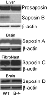
Saposin proteins in representative tissues of saposin B−/− mice. No saposin B was detected, whereas prosaposin was at WT levels in liver using anti-mouse saposin B antibody. Saposins A and D were at normal levels in B−/− brains detected with anti-mouse saposin A or saposin D antibodies, respectively. Using anti-mouse saposin C antibody, saposin C in B−/− fibroblast cells was slightly increased compared with WT controls.
Figure 3.
Figure 3.
Phenotypes of saposin B−/− mice. (A) Saposin B −/− mice exhibited hind-limb clasping during tail hanging by 15 months and (B) head tremor was evident at 22 months. (C) Narrow bridges test. B−/− mice were tested on 12 mm2 square beam. The latency (left) and foot slips (right) in B−/− became progressively different from WT with increasing age. Male mice were used in the tests. WT, n = 17; B−/−, n = 26. *P < 0.01; **P < 0.001; ***P = 0.0001 (Student's t-test).
Figure 4.
Figure 4.
Long-term potentiation (LTP) analyses of saposin B−/− hippocampal regions: LTP was recorded as the slope of resulting EPSPs from the parasagittal sections (350 µm) of hippocampal CA1 region. The slopes of the EPSP (LTP) were not significantly different between 15-month-old B−/− (black circles) and WT (open circles) male mice after recording for 90 min following stimulation. WT, n = 6; B−/−, n = 8.
Figure 5.
Figure 5.
Storage inclusions in neural tissues from B−/− mice: H&E staining of B−/− mice neural tissue sections showed that the inclusion materials (arrows) were present in neurons of spinal cord, brain stem, acoustic ganglion of inner ear and dorsal root ganglion. WT mice did not show inclusions in matched regions. The tissues sections were from 17-month-old WT and B−/− mice.
Figure 6.
Figure 6.
CD-68 and GFAP staining of CNS from B−/− mice. (Upper panel) Activated microglial cells were present in the CNS of B−/− mice. Microglial cells staining with anti-CD68 antibody (brown) in (A) WT and (B) B−/− cerebella, (C) B−/− spinal cord and (D) B−/− brain stem. The sections were counter stained with hematoxylin. (Lower panel) B−/− mice showed increased anti-GFAP (green) antibody positive cells indicating astrogliosis in several CNS regions. (E) WT and (F) B−/− thalami, (G) B−/− corpus colosum and (H) B−/− brain stem. The nuclei were stained with DAPI (blue). The tissues sections were from 17-month-old mice.
Figure 7.
Figure 7.
Alcian blue (sulfatide) staining in CNS of B−/− mice: (A) Alcian blue staining of B−/− tissues showed sulfatide (blue) storage in: spinal cord (A), kidney tubules (B), brain stem (E) and thalamus (F). WT spinal cord (C) and kidney (D) were free of alcian blue staining. (B) Consecutive spinal cord sections from B−/− mice were stained with alcian blue (A, C and E) and MBP (B), CD68 (D) and NeuN (F). (A and B) Alcian blue staining was localized to the MBP positive region (star). (C and D) CD68 positive microglial cells contained alcian blue positive material (arrows). (E and F) Alcian blue staining was on a neuronal process (arrow) and not in the neuronal soma. The tissues sections were from 15-month-old mice.
Figure 8.
Figure 8.
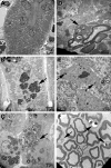
Ultrastructural kidney and CNS of saposin B−/− mice. (A) An electron micrograph of a normal proximal renal tubular cell. Rare and non specific small residual bodies were in the cytoplasm of the tubular lining cell. (B) Proximal renal tubular epithelial cells contained numerous large complex appearing multivesicular bodies (arrow). The storage materials consisted of aggregates of variable size heterogeneous granular material with slightly pale central cores surrounded by a rim of more electron dense amorphous material. (C) Renal epithelial cells from a distal tubule also contained a large amount of storage material (arrow). The material was similar to that seen in the proximal tubule in (B), although slightly less electron dense. A few of the vesicular bodies contained layers of membranous material surrounding less dense material in the central core. (D) An oligodendrocyte (arrow) in brain stem next to a myelinated process. The oligodendrocyte contained storage materials composed of dense membranous material surrounding more electron lucent material. (E) A neuron from brain stem had a few scattered inclusion bodies (arrows) representing storage materials. (F) A section through the sciatic nerve with myelinated fibers. The Schwann cells (arrows) contained inclusion bodies including large vacuoles mixed with electron dense amorphous material. The tissues were from 15-month-old mice.
Figure 9.
Figure 9.
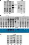
Glycosphingolipid analyses by TLC. (A) Lactosylceramide (LacCer, asterisk) was slightly increased in B−/− mice liver relative to WT, but lower than LacCer level in PS−/− mice (right panel). (B) Upper panel: Sulfatide (arrowheads) accumulated in B−/− kidney and was slightly elevated in B−/− lung. TriCer (arrows) was increased in B−/− kidney. Bottom panel: same plate as in the upper panel stained with Azure A to verify the sulfatide (arrowhead). (C) Gangliosides in B−/− mice cortex were not changed compared with WT cortex. WT and B−/− samples were from 15-month-old mice and PS−/− is from 4-week-old mice.
Figure 10.
Figure 10.
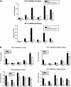
ESI/MS analysis of sulfatide. (A) Kidney. (Upper panel) Non-hydroxy (NFA) sulfatide C20, C22, C24 and C24:1 accumulated in B−/− mice compared with control samples. (Lower panel) Hydroxyl (HFA) sulfatides C20–C24:1 were increased relative to WT. Kidney samples were from 48-week-old mice. (B) Cortex and hippocampus. (Upper panel) NFA sulfatide C18, C24 and C24:1 were increased in B−/− mice. (Lower panel) HFA sulfatide C18-C24:1 were at higher levels than WT controls. Brain tissues were from 15-month-old mice. NFA sulfatide level was higher in cortex and hippocampus than kidney, whereas the HFA sulfatide level was greater in the kidney than brain sections. The amount of sulfatide were normalized by mg protein (n = 3).

References

    1. Rorman E.G., Scheinker V., Grabowski G.A. Structure and evolution of the human prosaposin chromosomal gene. Genomics. 1992;13:312–318. - PubMed
    1. O'Brien J.S., Kretz K.A., Dewji N., Wenger D.A., Esch F., Fluharty A.L. Coding of two sphingolipid activator proteins (SAP-1 and SAP-2) by same genetic locus. Science. 1988;241:1098–1101. - PubMed
    1. Collard M.W., Sylvester S.R., Tsuruta J.K., Griswold M.D. Biosynthesis and molecular cloning of sulfated glycoprotein 1 secreted by rat Sertoli cells: sequence similarity with the 70-kilodalton precursor to sulfatide/GM1 activator. Biochemistry. 1988;27:4557–4564. - PubMed
    1. Nakano T., Sandhoff K., Stumper J., Christomanou H., Suzuki K. Structure of full-length cDNA coding for sulfatide activator, a Co-beta-glucosidase and two other homologous proteins: two alternate forms of the sulfatide activator. J. Biochem (Tokyo) 1989;105:152–154. - PubMed
    1. Vielhaber G., Hurwitz R., Sandhoff K. Biosynthesis, processing, and targeting of sphingolipid activator protein (SAP)precursor in cultured human fibroblasts. Mannose 6-phosphate receptor-independent endocytosis of SAP precursor. J. Biol. Chem. 1996;271:32438–32446. - PubMed

Publication types