. 2008 May 20;105(20):7206-11.
doi: 10.1073/pnas.0802593105. Epub 2008 May 14.

Chaperone-dependent amyloid assembly protects cells from prion toxicity

Affiliations

Chaperone-dependent amyloid assembly protects cells from prion toxicity

Peter M Douglas et al. Proc Natl Acad Sci U S A. .

Abstract

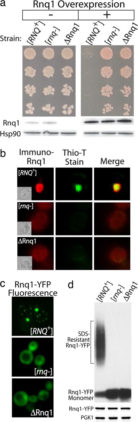
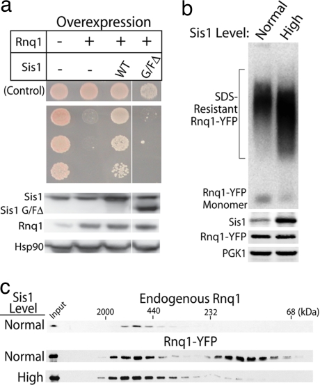
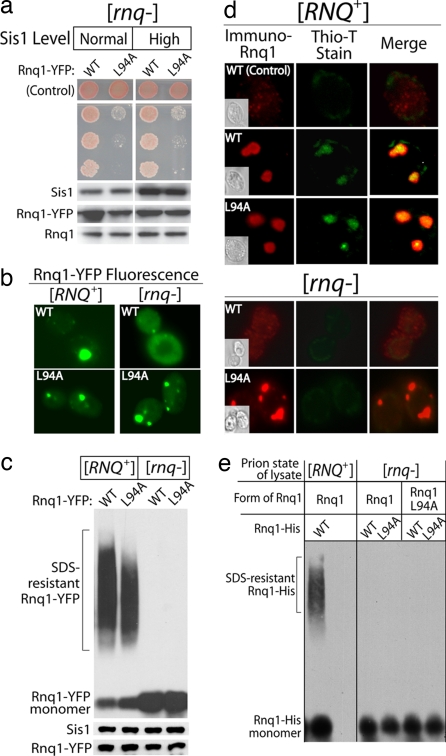
Protein conformational diseases are associated with the aberrant accumulation of amyloid protein aggregates, but whether amyloid formation is cytotoxic or protective is unclear. To address this issue, we investigated a normally benign amyloid formed by the yeast prion [RNQ(+)]. Surprisingly, modest overexpression of Rnq1 protein was deadly, but only when preexisting Rnq1 was in the [RNQ(+)] prion conformation. Molecular chaperones protect against protein aggregation diseases and are generally believed to do so by solubilizing their substrates. The Hsp40 chaperone, Sis1, suppressed Rnq1 proteotoxicity, but instead of blocking Rnq1 protein aggregation, it stimulated conversion of soluble Rnq1 to [RNQ(+)] amyloid. Furthermore, interference with Sis1-mediated [RNQ(+)] amyloid formation exacerbated Rnq1 toxicity. These and other data establish that even subtle changes in the folding homeostasis of an amyloidogenic protein can create a severe proteotoxic gain-of-function phenotype and that chaperone-mediated amyloid assembly can be cytoprotective. The possible relevance of these findings to other phenomena, including prion-driven neurodegenerative diseases and heterokaryon incompatibility in fungi, is discussed.


Conflict of interest statement

The authors declare no conflict of interest.

Figures

Fig. 1.
Fig. 1.
Overexpression of Rnq1 is toxic to [RNQ+] cells. (a) The effect of Rnq1 overexpression on yeast cell viability in the presence and absence of the [RNQ+] prion. (b) Thioflavin-T staining of Rnq1 in [RNQ+], [rnq−], and ΔRnq1 cells. Fixed yeast were decorated with α-Rnq1 sera that was detected with a fluorescent secondary antibody. The same cells were simultaneously stained with the amyloid indicator dye, thioflavin-T. (c) Visualization of the aggregation state of Rnq1-YFP by fluorescence microscopy. (d) (Upper) Assembly status of Rnq1-YFP as determined by SDD-AGE. (Lower) Western blots of cell extracts.
Fig. 2.
Fig. 2.
Sis1 overexpression protects against Rnq1 toxicity. (a) (Upper) The effect of Sis1 or Sis1ΔG/F overexpression on Rnq1 toxicity. Control indicates strains grown under noninducing conditions. (Lower) Western blots of the indicated proteins. (b) (Upper) Effect of Sis1 overexpression on the formation of SDS-resistant [RNQ+] conformers as determined by SDD-AGE. (Lower) Western blots of the indicated proteins. (c) Gel-filtration analysis of intracellular pools of endogenous Rnq1 versus overexpressed Rnq1-YFP.
Fig. 3.
Fig. 3.
Sis1 binding to a conserved chaperone-binding motif in the non-prion-forming domain of Rnq1. (a) A schematic showing the domain structure of Rnq1. The underlined region in the nonprion domain of Rnq1 represents a chaperone-binding motif identified via screening a cellulose peptide array (see Fig. S2). (b) Sis1-dependent binding of Hsp70 Ssa1 to the peptide in the Rnq1 peptide array that is bound most strongly by Sis1. (c) Mutation L94A in the chaperone-binding motif reduces the ability of Sis1 to form coimmunoprecipitable complexes with Rnq1-GFP in [RNQ+] cells. Rnq1-GFP was expressed by using the CUP1 promoter. Levels of the indicated proteins in c were visualized by Western blot (WB) analysis.
Fig. 4.
Fig. 4.
Mutations in the chaperone-binding motif of Rnq1 reduce the efficiency of [RNQ+] amyloid assembly. (a) Kinetics of Rnq1-GFP L94A assembly into SDS-resistant aggregates in [RNQ+] yeast determined by SDD-AGE. Rnq1-GFP fusions were expressed by using the CUP1 promoter. (b) [RNQ+] seed-dependent assembly of purified Rnq1-His and Rnq1-His L94A into SDS-resistant amyloid. (c) (Upper) Growth of 5-fold serial dilutions of [RNQ+] strains in which WT or L94A Rnq1 was overexpressed from the GAL1 promoter. Where indicated, Sis1 was overexpressed from the GPD promoter. (Lower) Relative expression level of the specified proteins as determined by Western blot.
Fig. 5.
Fig. 5.
Rnq1 L94A toxicity and assembly status in [rnq−] yeast. (a) (Upper) Growth of 5-fold serial dilutions of [rnq−] strains in which WT or L94A Rnq1 was overexpressed from the GAL1 promoter. Sis1 was overexpressed from the GPD promoter. (Lower) Western blots of cell extracts. (b) Fluorescent foci formed by Rnq1-YFP and Rnq1-YFP L94A in [RNQ+] and [rnq−] cells. (c) (Upper) SDD-AGE analysis of aggregates formed by Rnq1-YFP and Rnq1-YFP L94A in [RNQ+] and [rnq−] cells. (Lower) Western blots of cell extracts. (d) Thioflavin-T staining of untagged Rnq1 and Rnq1 L94A in [RNQ+] and [rnq−] cells. Fixed yeast was decorated with α-Rnq1 sera that was detected with a fluorescent secondary antibody. The same cells were simultaneously stained with the amyloid indicator dye, thioflavin-T. (e) Cell extracts from [rnq−] cells overexpressing either Rnq1 or Rnq1 L94A were incubated with purified Rnq1-His or Rnq1-His L94A. The assembly status of purified Rnq1-His was determined by SDD-AGE. As a control, the assembly status of purified Rnq1-His incubated with [RNQ+] cell extract also was determined.

References

    1. Carrell RW, Lomas DA. Conformational disease. Lancet. 1997;350:134–138. - PubMed
    1. Chiti F, Dobson CM. Protein misfolding, functional amyloid, and human disease. Annu Rev Biochem. 2006;75:333–366. - PubMed
    1. Caughey B, Lansbury PT. Protofibrils, pores, fibrils, and neurodegeneration: Separating the responsible protein aggregates from the innocent bystanders. Annu Rev Neurosci. 2003;26:267–298. - PubMed
    1. Nelson R, et al. Structure of the cross-beta spine of amyloid-like fibrils. Nature. 2005;435:773–778. - PMC - PubMed
    1. Haass C, Selkoe DJ. Soluble protein oligomers in neurodegeneration: Lessons from the Alzheimer's amyloid beta-peptide. Nat Rev Mol Cell Biol. 2007;8:101–112. - PubMed

Publication types