. 2008 May 14;28(20):5195-206.
doi: 10.1523/JNEUROSCI.1180-08.2008.

Gas6 deficiency increases oligodendrocyte loss and microglial activation in response to cuprizone-induced demyelination

Affiliations

Gas6 deficiency increases oligodendrocyte loss and microglial activation in response to cuprizone-induced demyelination

Michele D Binder et al. J Neurosci. .

Abstract

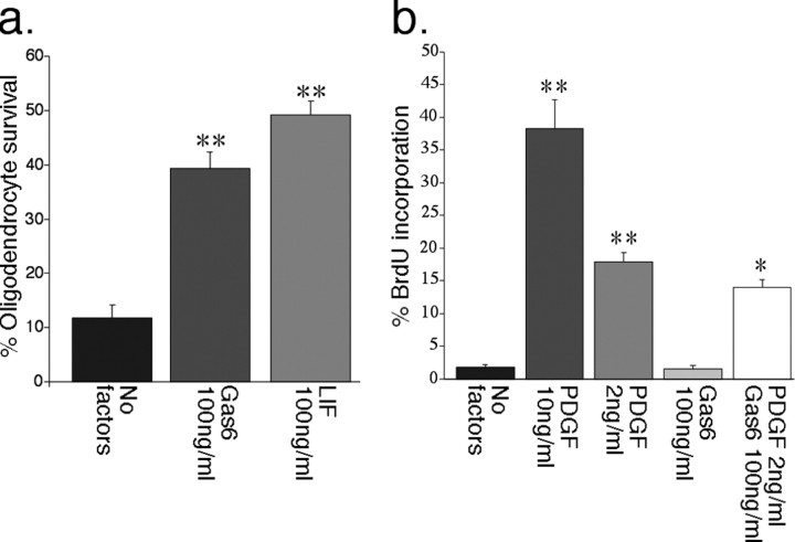
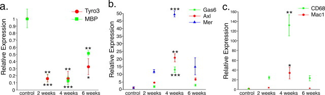
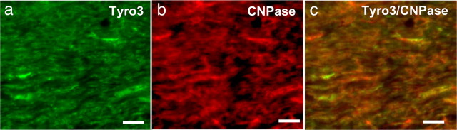
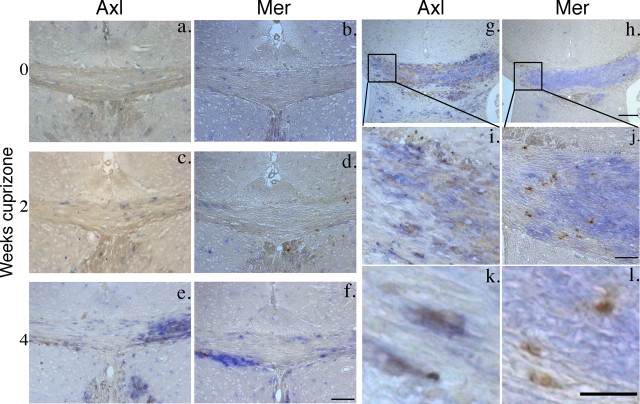
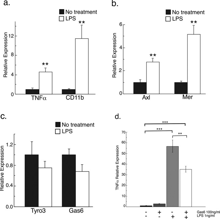
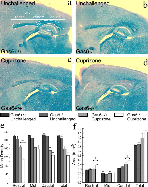
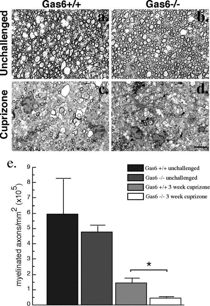
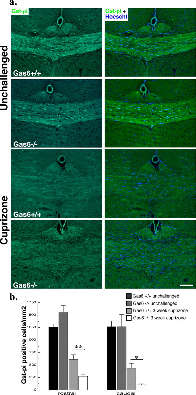
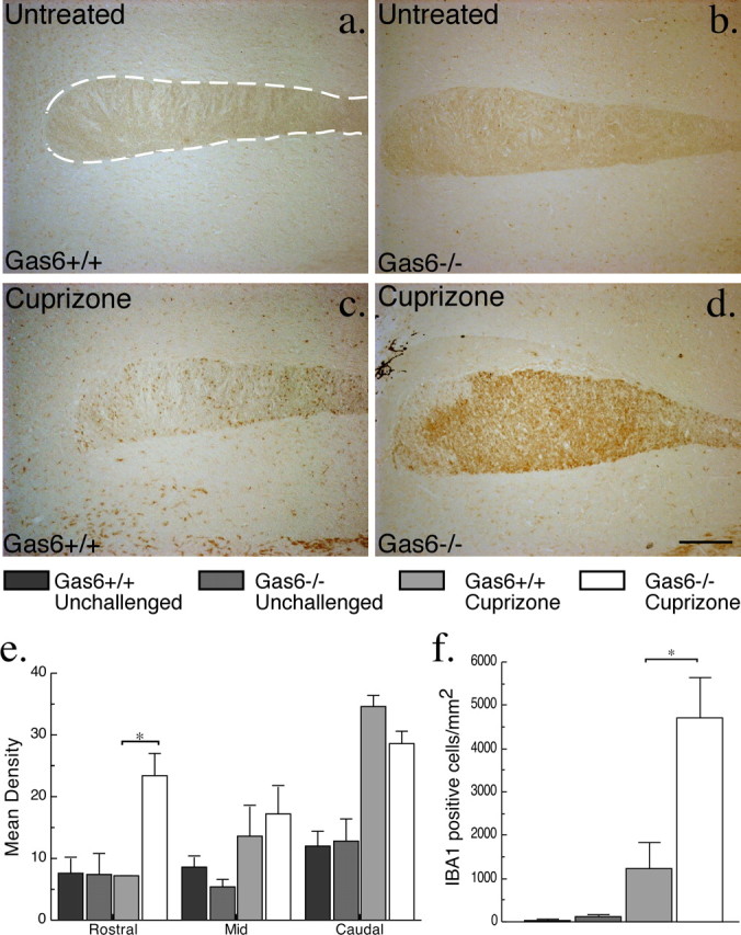
The TAM family of receptor protein tyrosine kinases comprises three known members, namely Tyro3, Axl, and Mer. These receptors are widely expressed in the nervous system, including by oligodendrocytes, the cell type responsible for myelinating the CNS. We examined the potential role of the TAM family and of their principle cognate ligand, Gas6 (growth arrest gene 6), in modulating the phenotype of the cuprizone model of demyelination. We found that the expression profiles of Axl, Mer, and Gas6 mRNA were increased in the corpus callosum in a temporal profile correlating with the increased migration and proliferation of microglia/macrophages in this model. In contrast, expression of Tyro3 decreased, correlating with the loss of oligodendrocytes. Gas6 both promoted in vitro survival of oligodendrocytes (39.3 +/- 3.1 vs 11.8 +/- 2.4%) and modulated markers of activation in purified cultures of microglia (tumor necrosis factor alpha mRNA expression was reduced approximately 48%). In Gas6-/- mice subjected to cuprizone-challenge, demyelination was greater than in control mice, within the rostral region of the corpus callosum, as assessed by luxol fast blue staining (myelination reduced by 36%) and by ultrastructural analysis. An increased loss of Gst-pi (glutathione S-transferase-pi)-positive oligodendrocytes was also identified throughout the corpus callosum of Gas6-/- mice. Microglial marker expression (ionized calcium-binding adapter molecule 1) was increased in Gas6-/- mice but was restricted to the rostral corpus callosum. Therefore, TAM receptor activation and regulation can independently influence both oligodendrocyte survival and the microglial response after CNS damage.


Figures

Figure 1.
Figure 1.
rhGas6 increases oligodendrocyte viability but not proliferation in vitro. a, Purified mature oligodendrocytes were plated at a density of 150 cells/well in SATO in the presence or absence of either rhGas6 or LIF. Cells were cultured for 48 h and then assessed for viability using calcein AM/ethidium homodimer-1. Six wells were counted per condition. Viability is expressed as a percentage of total cell number. Both LIF and Gas6 significantly increased the viability of cultured oligodendrocytes (**p < 0.001). b, Purified OPCs were plated at a density of 150 cells per well in SATO in the presence or absence of growth factors as specified and cultured for 48 h. BrdU was added for the final 17 h of culture. Cells were fixed and stained for BrdU, and the number of positive cells was counted. Results are expressed as a percentage of BrdU-positive cells per total cells counted (*p < 0.01, **p < 0.001, compared with no factors).
Figure 2.
Figure 2.
TAM receptor and Gas6 gene expression profiles are regulated during demyelination. a–c, The corpus callosum of unchallenged mice or mice challenged with cuprizone for 2, 4, or 6 weeks (n = 2 for each time point) was collected, and mRNA levels were measured using qPCR. a, Tyro3 and MBP mRNA expression profiles were significantly reduced at all time points examined (*p < 0.05, **p < 0.01, ***p < 0.001, compared with unchallenged mice). b, Gas6, Axl, and Mer mRNA expression profiles were significantly increased at 4 weeks of cuprizone challenge (**p < 0.01, ***p < 0.001). c, CD68 and CD11b mRNA expression profiles were significantly increased at 4 weeks of cuprizone challenge (*p < 0.05, **p < 0.01).
Figure 3.
Figure 3.
Tyro3 expression in oligodendrocytes of the corpus callosum. a, b, Coronal sections (15 μm) of 8 week mouse brains were double stained with anti-Tyro3 serum (a, green) and anti-CNPase (b, red) to label oligodendrocytes. c, Tyro3 was detected in oligodendrocytes as revealed by the yellow regions indicating overlap in the staining of Tyro3 and cell type-specific antibodies. Scale bar, 20 μm.
Figure 4.
Figure 4.
Microglia upregulate Axl and Mer in response to cuprizone challenge. a–f, Double-immunoperoxidase staining for Axl and Mer (brown) and IBA1 (blue) in the midline corpus callosum of unchallenged mice or mice challenged with cuprizone for 2 or 4 weeks (n = 4 for each time point). a, b, In unchallenged animals, a small number of IBA1-positive cells, evenly distributed throughout the corpus callosum, was observed. At this time point, cells positive for either Axl (a) or Mer (b) were rarely observed. c, d, After 2 weeks of cuprizone challenge, a small increase in the number of IBA1-positive cells was observed. k, l, Some of the IBA1-positive cells were also positive for Axl (k) or Mer (l). Cells positive for Axl or Mer alone were rarely observed. e, f, After 4 weeks of cuprizone challenge, the number of IBA1-positive cells in the corpus callosum was greatly increased, with a concomitant increase in Axl (e) and Mer (f) immunoreactivity among IBA1-positive cells, although Mer reactivity appeared less extensive. g, h, The dissociation between Axl- and Mer-positive cells is shown in staining of adjacent sections from mice challenged with cuprizone for 4 weeks. g–j, Axl positivity (g, i) appears to extend throughout the region of IBA1 expression, whereas Mer appears restricted to a subset of IBA1-positive microglia (h, j). Scale bars: a–f, i, j, 100 μm; g, h, 200 μm; k, l, 50 μm.
Figure 5.
Figure 5.
The expression profiles of TAM receptors and Gas6 mRNA are regulated in response to microglial activation, and activation is reduced in the presence of exogenous rhGas6. a–c, Purified rat microglia were incubated with or without 1 μg/ml LPS for 48 h, and mRNA was measured using qPCR. a, Levels of mRNA of both CDllb and TNFα, markers of microglial activation, increased in response to LPS treatment (**p < 0.01). b, Expression of both Axl and Mer mRNA increased in response to LPS-induced activation of microglia (**p < 0.01). c, In contrast, expression profiles of both Tyro3 and Gas6 mRNA in LPS-activated microglia were not significantly different from control cells (p > 0.05). d, Purified rat microglia were cultured in the presence or absence of rhGas6 for 3 h before activation was induced with LPS (1 ng/ml). Cells were harvested after 5 h of LPS treatment and RNA collected. Levels of TNFα mRNA, used as a surrogate marker for microglial activation, were measured by qPCR. There was a significant reduction in TNFα expression in rhGas6 treated microglia compared with microglia not treated with rhGas6 (**p < 0.01, ***p < 0.001).
Figure 6.
Figure 6.
Gas6−/− mice have decreased myelination and increased callosal area in response to cuprizone challenge. a–d, Wild-type and Gas6−/− mice were challenged with cuprizone for 3 weeks with age-matched unchallenged mice included as controls (n = 2, 3 mice per group). Sagittal sections were assessed using LFB staining with image analysis as described in Materials and Methods. e, A statistically significant decrease in LFB mean density was seen in the rostral segment of Gas6−/− animals challenged with cuprizone compared with similarly treated wild-type mice (*p < 0.05). f, The rostral segment also showed a statistically significant increase in area in Gas6−/− challenged with cuprizone (*p < 0.05). Additionally, there was a significant increase in area of the caudal segment of the corpus callosum in Gas6−/− mice (*p < 0.05). Scale bar, 100 μm. The dotted line indicates the boundaries of the corpus callosum (cc). ctx, Cortex; hc, hippocampus.
Figure 7.
Figure 7.
Gas6−/− mice have reduced numbers of myelinated axons in response to cuprizone challenge. Wild-type and Gas6−/− mice were challenged with cuprizone for 3 weeks with age-matched unchallenged mice included as controls (n = 2, 4 mice per group). a–d, Representative electron micrographs of the rostral segment of the corpus callosum. Unchallenged wild-type (a) and Gas6−/− (b) animals show a large number of healthy axons surrounded by compacted myelin. Cuprizone-challenged wild-type (c) and Gas6−/− (d) mice show reduced numbers of myelinated axons, with very few remaining in the Gas6−/− mice. Arrows indicate examples of myelinated axons, and arrowheads indicate examples of microglia with myelin debris. Scale bar, 2 μm. e, Semithin sections from within the rostral segment of the corpus callosum were stained with methylene blue, and the number of myelinated axons within this region was counted. No significant difference in the number of myelinated axons was seen in unchallenged mice (p > 0.05). In contrast, compared with their wild-type counterparts, Gas6−/− mice showed a significant reduction in the number of myelinated axons after cuprizone challenge (*p < 0.05).
Figure 8.
Figure 8.
Gas6−/− mice have decreased Gst-π-immunoreactive oligodendrocytes in response to cuprizone challenge. Wild-type and Gas6−/− mice were challenged with cuprizone for 3 weeks with age-matched unchallenged mice included as controls (n = 3, 5 for each group). Immunofluorescence analysis was performed to detect the presence of Gst-π reactive oligodendrocytes. a, Representative images of Gst-π staining and the same image merged with the nuclear stain Hoescht are shown. Scale bar, 100 μm. The number of Gst-π-positive cells per squared millimeter was determined as outlined in Materials and Methods, and the results are shown in b, with Gas6−/− mice showing a significant decrease in oligodendrocytes in both the rostral and caudal segments of the corpus callosum. *p < 0.05, **p < 0.01.
Figure 9.
Figure 9.
Gas6−/− mice have an increased number of IBA1-positive microglia/macrophages in the rostral corpus callosum in response to cuprizone challenge. Wild-type and Gas6−/− mice were challenged with cuprizone for 3 weeks with age-matched unchallenged mice included as controls (n = 2, 3 for each group). Immunohistochemistry was performed to detect the presence of IBA1 reactive microglia/macrophages. a–d, Representative images of the rostral segment of the corpus callosum are shown for each condition. The dotted line indicates the boundaries of the corpus callosum. Scale bar, 200 μm. The corpus callosum was divided into three separate segments, and the mean density of staining was determined as outlined in Materials and Methods. The results are shown in e, with Gas6−/− mice showing an increase in IBA1 staining density in the rostral segment (*p < 0.05). f, Adjacent sections were stained with IBA1 and counterstained with hemotoxylin to enable counting of individual cells in the rostral segment. No significant difference in the number of IBA1-positive cells was seen in unchallenged mice. In contrast, a significant increase in the number of IBA1-positive cells was observed in the cuprizone-challenged Gas6−/− mice compared with their wild-type counterparts (*p < 0.05).

Similar articles

Cited by

References

    1. Angelillo-Scherrer A, de Frutos P, Aparicio C, Melis E, Savi P, Lupu F, Arnout J, Dewerchin M, Hoylaerts M, Herbert J, Collen D, Dahlback B, Carmeliet P. Deficiency or inhibition of Gas6 causes platelet dysfunction and protects mice against thrombosis. Nat Med. 2001;7:215–221. - PubMed
    1. Arnett HA, Mason J, Marino M, Suzuki K, Matsushima GK, Ting JP. TNF alpha promotes proliferation of oligodendrocyte progenitors and remyelination. Nat Neurosci. 2001;4:1116–1122. - PubMed
    1. Arnett HA, Hellendall RP, Matsushima GK, Suzuki K, Laubach VE, Sherman P, Ting JP. The protective role of nitric oxide in a neurotoxicant-induced demyelinating model. J Immunol. 2002;168:427–433. - PubMed
    1. Arnett HA, Wang Y, Matsushima GK, Suzuki K, Ting JP. Functional genomic analysis of remyelination reveals importance of inflammation in oligodendrocyte regeneration. J Neurosci. 2003;23:9824–9832. - PMC - PubMed
    1. Barnett MH, Prineas JW. Relapsing and remitting multiple sclerosis: pathology of the newly forming lesion. Ann Neurol. 2004;55:458–468. - PubMed

Publication types

MeSH terms