. 2008 Aug;82(15):7640-52.
doi: 10.1128/JVI.00170-08. Epub 2008 May 14.

A role for E1B-AP5 in ATR signaling pathways during adenovirus infection

Affiliations

A role for E1B-AP5 in ATR signaling pathways during adenovirus infection

Andrew N Blackford et al. J Virol. 2008 Aug.

Abstract

E1B-55K-associated protein 5 (E1B-AP5) is a cellular, heterogeneous nuclear ribonucleoprotein that is targeted by adenovirus (Ad) E1B-55K during infection. The function of E1B-AP5 during infection, however, remains largely unknown. Given the role of E1B-55K targets in the DNA damage response, we examined whether E1B-AP5 function was integral to these pathways. Here, we show a novel role for E1B-AP5 as a key regulator of ATR signaling pathways activated during Ad infection. E1B-AP5 is recruited to viral replication centers during infection, where it colocalizes with ATR-interacting protein (ATRIP) and the ATR substrate replication protein A 32 (RPA32). Indeed, E1B-AP5 associates with ATRIP and RPA complex component RPA70 in both uninfected and Ad-infected cells. Additionally, glutathione S-transferase pull-downs show that E1B-AP5 associates with RPA components RPA70 and RPA32 directly in vitro. E1B-AP5 is required for the ATR-dependent phosphorylation of RPA32 during infection and contributes to the Ad-induced phosphorylation of Smc1 and H2AX. In this regard, it is interesting that Ad5 and Ad12 differentially promote the phosphorylation of RPA32, Rad9, and Smc1 during infection such that Ad12 promotes a significant phosphorylation of RPA32 and Rad9, whereas Ad5 only weakly promotes RPA32 phosphorylation and does not induce Rad9 phosphorylation. These data suggest that Ad5 and Ad12 have evolved different strategies to regulate DNA damage signaling pathways during infection in order to promote viral replication. Taken together, our results define a role for E1B-AP5 in ATR signaling pathways activated during infection. This might have broader implications for the regulation of ATR activity during cellular DNA replication or in response to DNA damage.


Figures

FIG. 1.
FIG. 1.
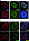
Effects of Ad infection on protein levels of E1B-55K binding proteins. A549 cells were infected with 10 PFU per cell of either wt Ad5 (A) or wt Ad12 (B). Cells were harvested at the appropriate times postinfection, and 50 μg protein samples was separated by SDS-PAGE and transferred onto nitrocellulose. Membranes were then Western blotted for E1B-AP5 (i), MRE11 (ii), p53 (iii), E1B-55K (iv), and β-actin (v) using the appropriate antibodies and visualized by enhanced chemiluminescence. (C) Immunofluorescent Zeiss LSM510-Meta confocal microscopic detection of E1B-AP5 protein expression in wt Ad5- and wt Ad12-infected A549 cells. Cells were infected with either 10 PFU/cell wt Ad5 or wt Ad12, subsequently fixed with 4% (wt/vol) paraformaldehyde in PBS, and permeabilized with acetone 24 h postinfection. E1B-AP5 (i and iv), Ad5 DBP (ii), and Ad12 E1B-55K (v) were then visualized using the appropriate antibodies. Nuclei are stained with DAPI and are shown in blue.
FIG. 2.
FIG. 2.
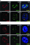
(A) Localization of E1B-AP5, ATRIP, and RPA32 in mock-infected interphase A549 cells. Cells were grown on glass coverslips and then treated with preextraction buffer and fixed with 4% (wt/vol) paraformaldehyde as described in Materials and Methods. Antigens were detected using the appropriate antibodies. (B) Localization of pTP, DBP, and RPA32 at viral replication centers in Ad5-infected A549 cells. Twenty-four hours postinfection with 10 PFU/cell wt Ad5, cells were treated with preextraction buffer and then fixed with 4% (wt/vol) paraformaldehyde, whereupon antigens were detected using the appropriate reagents. Colocalization images were recorded using a Zeiss LSM510-Meta laser scanning confocal microscope. Nuclei are stained with DAPI and are shown in blue.
FIG. 3.
FIG. 3.
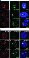
Colocalization of E1B-AP5 and RPA32, and ATRIP and RPA32, at Ad5 (A) and Ad12 (B) replication centers. Cells were infected with 10 PFU/cell of the appropriate virus. Ad5-infected and Ad12-infected cells were treated with preextraction buffer and then fixed with 4% (wt/vol) paraformaldehyde 24 h and 48 h postinfection, respectively. E1B-AP5, RPA32, and ATRIP localizations were visualized using the appropriate antibodies. Colocalization images were recorded using a Zeiss LSM510-Meta confocal microscope. Regions of substantial colocalization are shown as yellow in the merged image. Nuclei are stained with DAPI and are shown in blue.
FIG. 4.
FIG. 4.
(A) Colocalization of E1B-AP5 with E1B-55K at Ad5 and Ad12 replication centers. Cells were infected with 10 PFU/cell of the appropriate virus. Infected cells were treated with preextraction buffer and then fixed with 4% (wt/vol) paraformaldehyde 24 h postinfection. E1B-AP5 and Ad E1B-55K were visualized using the appropriate antibodies. (B) Recruitment of E1B-AP5, RPA32, and ATRIP to Ad5 replication centers is independent of E1B-55K. Cells were infected with 10 PFU/cell of Ad5 dl1520 and treated with preextraction buffer and then fixed with 4% (wt/vol) paraformaldehyde 24 h postinfection. E1B-AP5, Ad5 DBP, RPA32, and ATRIP were visualized using the appropriate antibodies. Colocalization images were recorded using a Zeiss LSM510-Meta confocal microscope. Regions of substantial colocalization are shown as yellow in the merged image. Nuclei are stained with DAPI and are shown in blue.
FIG. 5.
FIG. 5.
(A and B) Association of E1B-AP5 with ATRIP, RPA70, and DBP in uninfected and Ad5-infected A549 cells. Asynchronously growing, mock-infected, and Ad5-infected (10 PFU/cell) A549 cells were harvested 24 h postinfection and subjected to immunoprecipitation with either anti-IgG, -SAF-B, or -E1B-AP5 antibodies. Immunoprecipitates were collected on protein G-Sepharose beads, eluted with the appropriate sample buffer, separated by SDS-PAGE, and transferred onto nitrocellulose. ATM, ATRIP, RPA70, and DBP were visualized by Western blotting with the appropriate antibodies. (C) Association of E1B-AP5 with the RPA components RPA32 and RPA70 in vitro. Ten micrograms of GST-E1B-AP5 fusion protein was incubated with 20 μl of l-α-[35S]methionine-labeled RPA70, RPA32, or RPA14. Bound proteins were precipitated using glutathione-Sepharose and selectively eluted with glutathione (see Materials and Methods). Proteins were separated by SDS-PAGE and subjected to fluorography (Amersham Pharmacia). Gels were dried and then subjected to autoradiography.
FIG. 6.
FIG. 6.
Colocalization of Rad9, Rad17, and RPA32 at Ad replication centers. A549 cells were either mock infected (A) or infected with 10 PFU/cell of wt Ad5 (Bi to iii and Ci to iii), wt Ad12 (Biv to vi and Civ to vi), or Ad5 dl1520 (Di to vi). Ad5- and Ad12-infected cells were treated with preextraction buffer and then fixed with 4% (wt/vol) paraformaldehyde at 24 h and 48 h postinfection, respectively. Rad9, Rad17, and RPA32 were visualized using the appropriate antibodies. Colocalization images were recorded using a Zeiss LSM510-Meta confocal microscope. Regions of substantial colocalization are shown in yellow in the merged image. Nuclei are stained with DAPI and are shown in blue.
FIG. 7.
FIG. 7.
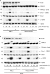
(A) Ad5 and Ad12 differentially regulate the phosphorylation of RPA32, Rad9, and Smc1 during infection. A549 cells were infected with 10 PFU/cell of either wt Ad5 or wt Ad12. Cells were harvested at the appropriate times postinfection, and 50 μg protein samples was separated by SDS-PAGE. After electrophoretic transfer onto nitrocellulose, membranes were Western blotted for RPA32 (i), RPA32 S4/8 (ii), Rad9 (iii), Smc1-S966 (iv), Smc1 (v), Chk1-S345 (vi), Chk1 (vii), γ-H2AX (viii), and H2AX (ix) using the appropriate antibodies. (B) E1B-AP5 is required for Ad12-induced phosphorylation of RPA32. A549 cells were initially treated with either nonsilencing (non-sil.) siRNA or siRNA oligonucleotides specific for the E1B-AP5 gene. Cells were subsequently infected with either Ad5 or Ad12 (at 10 PFU/cell), and whole-cell lysates were prepared at the appropriate times postinfection. After SDS-PAGE and transfer onto nitrocellulose, membranes were probed for E1B-AP5 (i), RPA32 (ii), RPA32 S4/8 (iii), Smc1-S966 (iv), Smc1 (v), γ-H2AX (vi), and H2AX (vii) with the appropriate antibodies. Antigens were visualized by ECL.
FIG. 8.
FIG. 8.
Role of ATM and other caffeine-sensitive kinases in Ad5 (A)- and Ad12 (B)-induced phosphorylation events. HeLa cells and HeLa shATM cells were infected with either Ad5 or Ad12 (at 10 PFU/cell), following which they were incubated in the absence or presence of 5 mM caffeine. Cells were harvested at the appropriate times postinfection, and 50 μg protein samples was separated by SDS-PAGE. After electrophoretic transfer onto nitrocellulose, membranes were Western blotted for ATM (i), RPA32 (ii), RPA32 S4/8 (iii), Smc1-S966 (iv), Smc1 (v), γ-H2AX (vi), and H2AX (vii) using the appropriate antibodies and visualized by ECL.
FIG. 9.
FIG. 9.
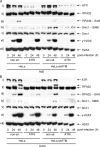
Role of ATR in Ad5 (A)- and Ad12 (B)-induced phosphorylation events. HeLa cells and HeLa shATM cells were initially treated with either nonsilencing (non-sil.) siRNA or siRNA oligonucleotides specific for the ATR gene. Cells were subsequently infected with either Ad5 or Ad12 (at 10 PFU/cell). Cells were harvested at the appropriate times postinfection, and 50 μg protein samples was separated by SDS-PAGE. After electrophoretic transfer onto nitrocellulose, membranes were Western blotted for ATR (i), RPA32 (ii), RPA32 S4/8 (iii), Smc1-S966 (iv), Smc1 (v), γ-H2AX (vi), and H2AX (vii) using the appropriate antibodies and visualized by ECL.

References

    1. Araujo, F. D., T. H. Stracker, C. T. Carson, D. V. Lee, and M. D. Weitzman. 2005. Adenovirus type 5 E4orf3 protein targets the Mre11 complex to cytoplasmic aggresomes. J. Virol. 7911382-11391. - PMC - PubMed
    1. Baker, A., K. J. Rohleder, L. A. Hanakahi, and G. Ketner. 2007. Adenovirus E4 34k and E1b 55k oncoproteins target host DNA ligase IV for proteasomal degradation. J. Virol. 817034-7040. - PMC - PubMed
    1. Barral, P. M., A. Rusch, A. S. Turnell, P. H. Gallimore, P. J. Byrd, T. Dobner, and R. J. A. Grand. 2005. The interaction of the hnRNP family member E1B-AP5 with p53. FEBS Lett. 5792752-2758. - PubMed
    1. Berk, A. J. 2005. Recent lessons in gene expression, cell cycle control, and cell biology from adenovirus. Oncogene 247673-7685. - PubMed
    1. Binz, S. K., A. M. Sheehan, and M. S. Wold. 2004. Replication protein A phosphorylation and the cellular response to DNA damage. DNA Repair 31015-1024. - PubMed

Publication types

MeSH terms

Substances