. 2008 Jul;82(14):7078-88.
doi: 10.1128/JVI.00619-08. Epub 2008 May 14.

The immune response to herpes simplex virus type 1 infection in susceptible mice is a major cause of central nervous system pathology resulting in fatal encephalitis

Affiliations

The immune response to herpes simplex virus type 1 infection in susceptible mice is a major cause of central nervous system pathology resulting in fatal encephalitis

Patric Lundberg et al. J Virol. 2008 Jul.

Abstract

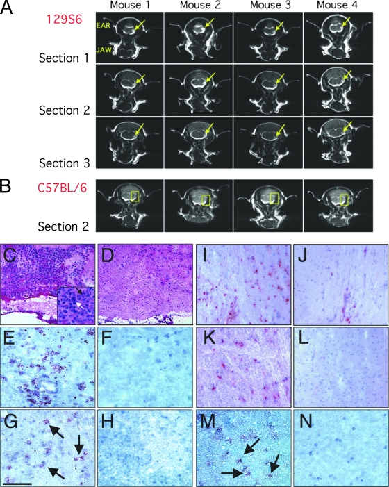
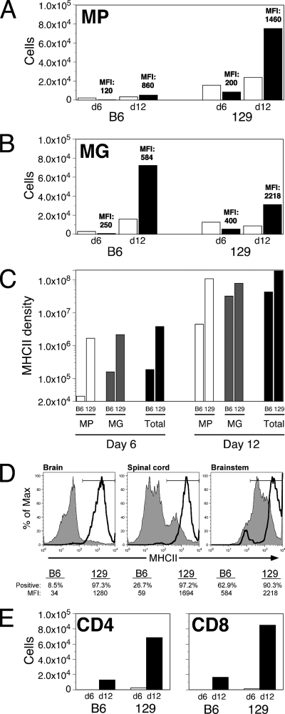
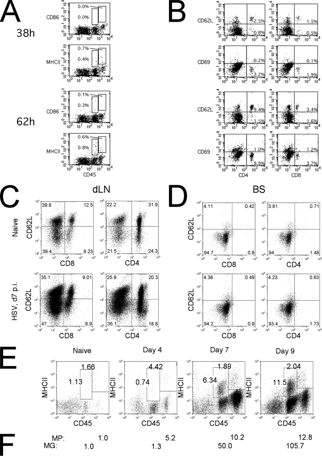
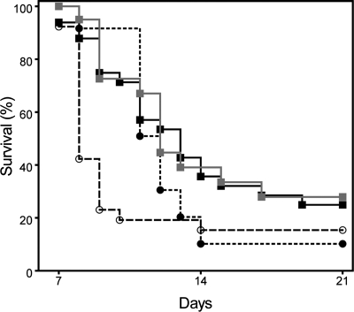
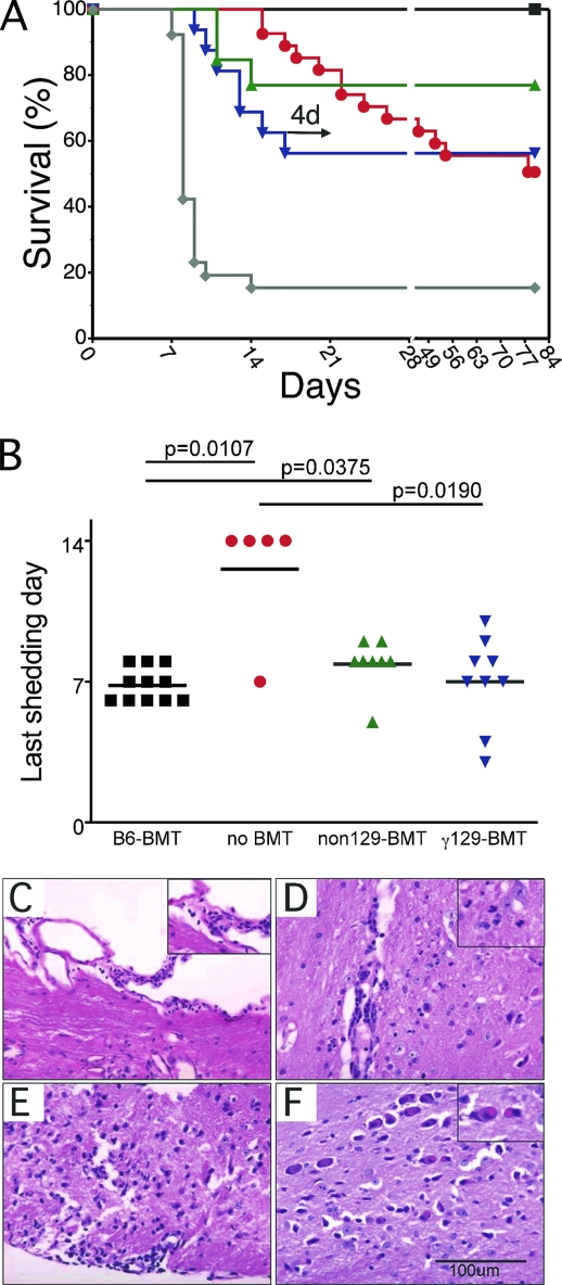
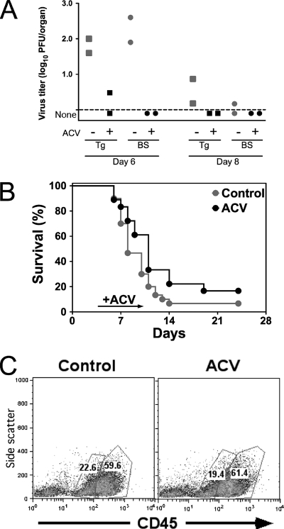
This study was undertaken to investigate possible immune mechanisms in fatal herpes simplex virus type 1 (HSV-1) encephalitis (HSE) after HSV-1 corneal inoculation. Susceptible 129S6 (129) but not resistant C57BL/6 (B6) mice developed intense focal inflammatory brain stem lesions of primarily F4/80(+) macrophages and Gr-1(+) neutrophils detectable by magnetic resonance imaging as early as day 6 postinfection (p.i.). Depletion of macrophages and neutrophils significantly enhanced the survival of infected 129 mice. Immunodeficient B6 (IL-7R(-/-) Kit(w41/w41)) mice lacking adaptive cells (B6-E mice) and transplanted with 129 bone marrow showed significantly accelerated fatal HSE compared to B6-E mice transplanted with B6 marrow or control nontransplanted B6-E mice. In contrast, there was no difference in ocular viral shedding in B6-E mice transplanted with 129 or B6 bone marrow. Acyclovir treatment of 129 mice beginning on day 4 p.i. (24 h after HSV-1 first reaches the brain stem) reduced nervous system viral titers to undetectable levels but did not alter brain stem inflammation or mortality. We conclude that fatal HSE in 129 mice results from widespread damage in the brain stem caused by destructive inflammatory responses initiated early in infection by massive infiltration of innate cells.


Figures

FIG. 1.
FIG. 1.
Visualization of BS lesions by MRI and histology from HSV-infected 129 and B6 mice. (A and B) Coronal T2-weighted MRIs at the level of the trigeminal nerve root in four 129 mice (A) (three consecutive MRI sections, with arrows indicating increased signals in trigeminal nuclei) and four B6 mice (B) (corresponding to section 2 in panel A, with rectangles outlining the areas of the trigeminal nuclei not showing increased signals) at day 6 p.i. Data shown are representative of two experiments. (C and D) H&E-stained, frozen BS sections from an infected 129 mouse (C) (with an extensive inflammatory infiltrate [shown at a higher magnification in the inset] composed of neutrophils [black arrow] and mononuclear cells [white arrow]) and a B6 mouse (D) (the corresponding section with scant inflammatory cells) at day 7 p.i. (E to M) BS immunohistochemical staining at day 7 p.i. of 129 mice positive for perivascular F4/80+ macrophages (E), Gr-1+ neutrophils (G), CD4+ T cells (I), CD8+ T cells (K), and HSV antigen (M), with corresponding negative immunochemical staining in B6 mice (F, H, L, M), except for an occasional perivascular T cell, as shown in panel J. The bar shown in panel G is 75 μm.
FIG. 2.
FIG. 2.
Accumulation and activation of immune cells over time in BS of susceptible and resistant mice. Cells were isolated from infected BS of B6 and 129 mice on day 6 or day 12 p.i. and analyzed for surface marker expression. (A) Macrophages (MP; CD11b+ F4/80+ CD45hi). (B) Microglia (MG; F4/80low CD45int). (C) Bar graphs indicate the absolute cell numbers of MHC-II-negative (open bars) and -positive (filled bars) macrophages recovered from pooled BS (n = 3). MFI is indicated for the MHC-II+ gates. Bar graphs of total MHC-II density on macrophages (white bars), microglia (gray bars), and MP plus MG (Total; black bars) in the BS of B6 (left) and 129 (right) mice on days 6 and 12 are shown. MHC-II density was calculated by multiplying the absolute number of MHC-II+ cells by their MFIs, as indicated in panels A and B, and provides a measure of the relative number of MHC molecules in the BS. (D) MHC-II expression on MG in three different CNS compartments of B6 (filled gray histograms) and 129 (open thick line) mice on day 12. The MFI is also indicated for each strain. MG were gated as viable forward scatter-side scatter and side scatter-CD45int events, and data represent typical population profiles from pooled tissues of two to four mice and are representative of two experiments. The MHC-II+ gate is shown for each histogram overlay, and the percentage of positive cells for each strain and tissue is given below each panel. (E) Absolute numbers of CD4+ CD45hi (left panel) or CD8+ CD45hi (right panel) T-cells in BS from B6 and 129 mice on days 6 (open bars) and 12 (filled bars).
FIG. 3.
FIG. 3.
Infiltrating cells in the BS of HSV-infected 129 mice and activation of microglia. Cells isolated from BS of HSV-infected 129 mice at 38 h and 62 h p.i. were stained for CD45 (A) and CD4 and CD8 (B) surface markers and activation molecules (MHC-II or CD86 [A] and CD62L or CD69 [B]). Increases in numbers and activation (loss of CD62L) of CD4 and CD8 T cells in dLNs on day 7 (C) were not seen for CD4 and CD8 T cells retrieved from BS of the same mice (D). BS cells stained for CD45 and MHC-II showed a continued increase in activation of microglia (CD45int) until day 9 p.i. (with peaks at 11.5% of the total). (E) Absolute increases in macrophages (MP) and microglias (MG) relative to levels for naïve mice (day 0) are indicated below panel E for days 4 to 9 p.i. (F) Data shown are representative of three experiments for panels A and B and two experiments for panels C, D, and E.
FIG. 4.
FIG. 4.
Depletion of neutrophils or macrophages during acute HSV infection enhances survival. Compared to mock treatment of 129 mice (open circles, dashed line, PBS or rat immunoglobulin G), Gr-1 MAb (black squares), clodronate (gray squares), and combination treatment with both Gr-1 MAb and clodronate (black circles) significantly delayed mortality (the mean survival time was prolonged to day 12 or 13, versus day 8 for controls). Survival on day 10 was highly significant for all treatment groups (P < 0.003 for each pairwise comparison to 129 mock controls). Data shown are combined from five different experiments. The overall statistical differences at the end of the experiments were as indicated: **, P < 0.005; *, P < 0.05 for comparison to the MOCK group; ns, not significantly different.
FIG. 5.
FIG. 5.
Transfer of susceptibility and resistance to fatal HSE by BMT. (A) B6-E mice were given no transplant (red circles), given B6 bone marrow (black squares), γ irradiated and given 129 bone marrow (inverted blue triangles), or nonirradiated and given 129 bone marrow (green triangles). Data for 129S6 mice are indicated for comparison (gray diamonds). As indicated by the arrow, 4 days (4d) after the last γ-irradiated mouse receiving 129 BMT died, >80% of non-BMT mice were still alive (P = 0.026 [significant]). Mice were infected by corneal scarification with 3,200 PFU HSV-1 17+, and mortality was monitored. Data shown are combined from four experiments. (B) Duration of shedding for individual mice from different groups (as indicated in the figure) is plotted, with mean time of last day of shedding (horizontal line) indicated. Horizontal bars above the graph indicate statistically significant differences between connected groups (representative of two experiments). BS from all four groups of BMT recipients in panel A were examined for pathology associated with HSV infection. Occasional or no viral inclusions were found in neurons in controls receiving no transplant and in recipients of B6 BMT (not shown). The abnormalities found in nonirradiated recipients of 129 BMT were less severe than those in their γ-irradiated counterparts. Shown are sections from γ-irradiated mice receiving 129 BMT. Cellular infiltrates are present in the subarachnoid space (C), perivascular parenchymal infiltrates in the BS (D) (neutrophils are shown in the inset), subpial infiltrating inflammatory cells (E), and prominent viral inclusions in the trigeminal nucleus (F). The inset shows higher magnification of several neurons with nuclear inclusions.
FIG. 6.
FIG. 6.
Effect of ACV on nervous system HSV titer, mortality, and BS inflammation. 129 mice were inoculated with HSV by corneal scarification and given daily i.p. injections with PBS (gray symbols) or 50 mg/kg ACV (black symbols) from day 4 to 10 p.i. (A) HSV titers of trigeminal ganglia (squares) and BS (circles) (data shown are from one of two trials, with two mice in each group sacrificed at days 6 and 8 p.i.); (B) mortality due to HSE (20 control and 14 ACV-treated mice); (C) BS-infiltrating mononuclear cells at day 10 p.i. (CD45high cells are peripheral leukocytes, while CD45int cells are CD11b+ microglia).

Similar articles

Cited by

References

    1. Andrews, D. M., V. B. Matthews, L. M. Sammels, A. C. Carrello, and P. C. McMinn. 1999. The severity of Murray Valley encephalitis in mice is linked to neutrophil infiltration and inducible nitric oxide synthase activity in the central nervous system. J. Virol. 738781-8790. - PMC - PubMed
    1. Aravalli, R. N., S. Hu, T. N. Rowen, J. M. Palmquist, and J. R. Lokensgard. 2005. Cutting edge: TLR2-mediated proinflammatory cytokine and chemokine production by microglial cells in response to herpes simplex virus. J. Immunol. 1754189-4193. - PubMed
    1. Bale, J. F., and L. J. Miner. 2005. Herpes simplex virus infections of the newborn. Curr. Treat. Options Neurol. 7151-156. - PubMed
    1. Cantin, E. M., D. R. Hinton, J. Chen, and H. Openshaw. 1995. Gamma interferon expression during acute and latent nervous system infection by herpes simplex virus type 1. J. Virol. 694898-4905. - PMC - PubMed
    1. Chen, S.-H., H.-W. Yao, W.-Y. Huang, K.-S. Hsu, H.-Y. Lei, A.-L. Shiau, and S.-H. Chen. 2006. Efficient reactivation of latent herpes simplex virus from mouse central nervous system tissues. J. Virol. 8012387-12392. - PMC - PubMed

Publication types

MeSH terms