. 2008 Jul;82(14):7155-66.
doi: 10.1128/JVI.02543-07. Epub 2008 May 14.

CDK13, a new potential human immunodeficiency virus type 1 inhibitory factor regulating viral mRNA splicing

Affiliations

CDK13, a new potential human immunodeficiency virus type 1 inhibitory factor regulating viral mRNA splicing

Reem Berro et al. J Virol. 2008 Jul.

Abstract

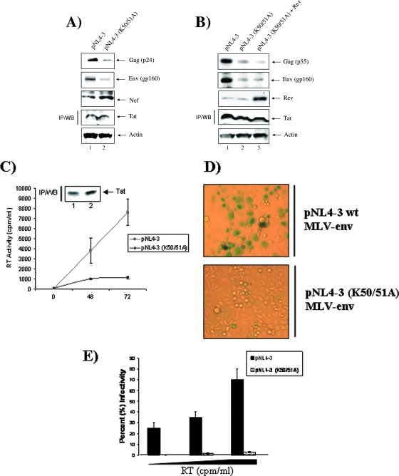
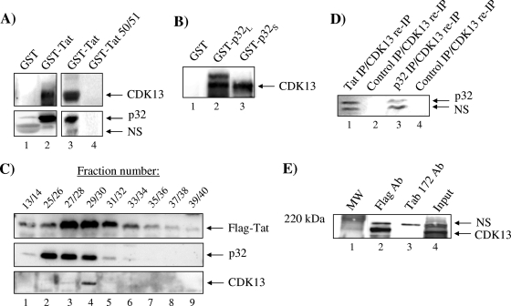
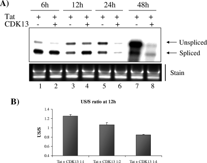
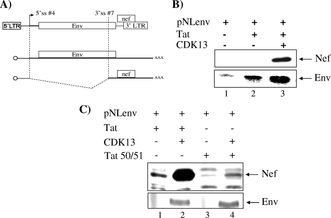
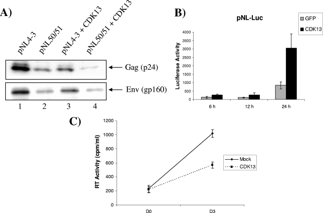
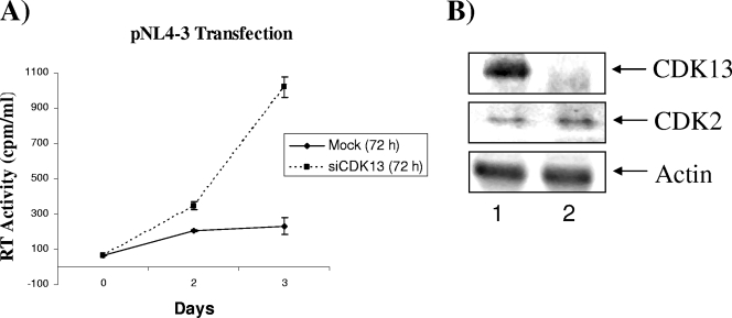
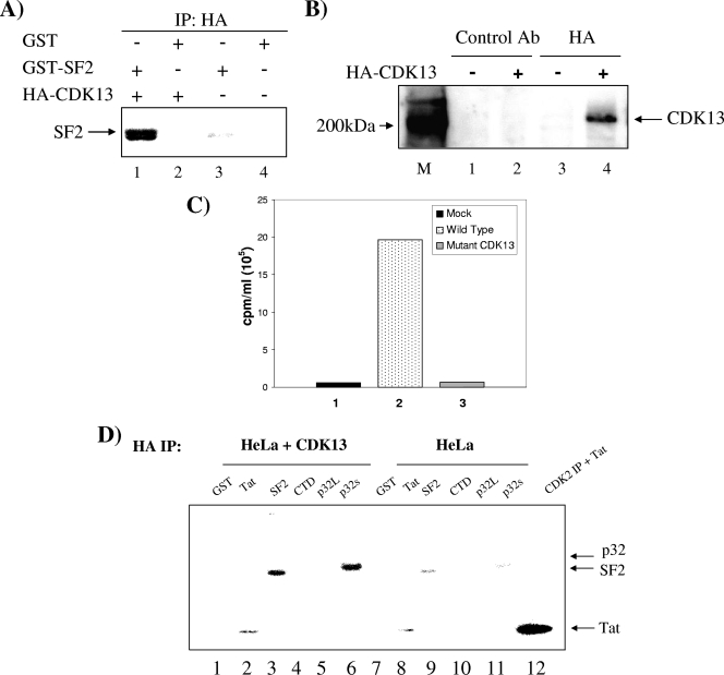
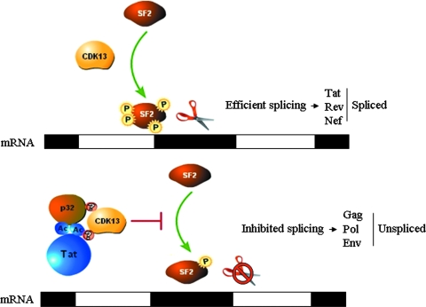
The human immunodeficiency virus type 1 (HIV-1) Tat is a 14-kDa viral protein that acts as a potent transactivator by binding to the transactivation-responsive region, a structured RNA element located at the 5' end of all HIV-1 transcripts. Tat transactivates viral gene expression by inducing the phosphorylation of the C-terminal domain of RNA polymerase II through several Tat-activated kinases and by recruiting chromatin-remodeling complexes and histone-modifying enzymes to the HIV-1 long terminal repeat. Histone acetyltransferases, including p300 and hGCN5, not only acetylate histones but also acetylate Tat at lysine positions 50 and 51 in the arginine-rich motif. Acetylated Tat at positions 50 and 51 interacts with a specialized protein module, the bromodomain, and recruits novel factors having this particular domain, such as P/CAF and SWI/SNF. In addition to having its effect on transcription, Tat has been shown to be involved in splicing. In this study, we demonstrate that Tat interacts with cyclin-dependent kinase 13 (CDK13) both in vivo and in vitro. We also found that CDK13 increases HIV-1 mRNA splicing and favors the production of the doubly spliced protein Nef. In addition, we demonstrate that CDK13 acts as a possible restriction factor, in that its overexpression decreases the production of the viral proteins Gag and Env and subsequently suppresses virus production. Using small interfering RNA against CDK13, we show that silencing of CDK13 leads to a significant increase in virus production. Finally, we demonstrate that CDK13 mediates its effect on splicing through the phosphorylation of ASF/SF2.


Figures

FIG. 1.
FIG. 1.
Tat acetylation mutant has reduced virus production. (A and B) Western blotting (WB) to detect the different viral proteins by use of antibodies against Gag (1:200), Env (1:200), and Nef (1:1,000) in 293T cells transfected with pNL4-3 or pNL4-3 (K50, 51A) for 48 h in the absence (A) or presence (B) of Rev. (C) RT assay to determine virus production in supernatant of 293T cells transfected with pNL4-3 or pNL4-3 (K50, 51A) collected at 0, 48, and 72 h after transfection. The data are representative of three independent experiments. (Inset) Similar amounts of Tat were seen from both wt and mutant clones after 72 h. (D) Magi cell assay to determine infectivity of viruses harvested from 293T cells transfected with pNL4-3 or pNL4-3 (K50, 51A) collected 72 h after transfection. Magi cells were infected with the wt and mutant viruses. Blue cells are infected cells. (E) Infectivity was determined by calculating the percentage of infected cells (blue cells) in the Magi cell assay system after infection for 48 h with increasing amounts of virus and with increasing RT activity. The data are representative of three independent experiments.
FIG. 2.
FIG. 2.
CDK13 and Tat interact both in vitro and in vivo. (A) GST-Tat or GST (1 μg) was incubated in HeLa cell extract (1 mg) in order to pull down CDK13. Glutathione beads were added and then the precipitated complex was washed and separated on a 4 to 20% Tris-glycine polyacrylamide gel. Western blotting was then performed using CDK13 (1:1,000), p32 (1:1,000), and U1 70K (1:200) antibodies to determine whether these proteins were pulled down by GST-Tat. A control experiment utilized GST-Tat 50/51 (1 μg) (lane 3). (B) GST-p32 was used to determine whether p32 binds CDK13 under conditions similar to those described for panel A. (C) 293T cells lysated from the cells infected with adeno-Tat were fractionated using a Sepharose 6 chromatography column and analyzed with indicated antibodies by immunoblotting (p32 and CDK13) or IP (Flag). (D) IP followed by re-IP was done with pulled fractions of 27 to 31 (100 μl, ∼50 μg of protein) and anti-Tat (polyclonal immunoglobulin G [IgG]-purified Ab, 5 μg) (lane 1), preimmune IgG sera (5 μg) (lane 2), anti-p32 (∼1 μg) (lane 3), or IgG purified (1 μg) overnight with TNE50 plus 0.1% NP-40 (lane 4). The next day, samples were pulled down with protein A/G beads, washed with TNE150, and eluted with 50 μl of radioimmunoprecipitation assay buffer. Eluates were re-IPed with CDK13 (∼1 μg) and Western blotted with anti-p32 Ab. (E) HLM-1 cells were transfected with Flag-Tat plasmid, and Flag Ab (5 μg) was used to pull down the Flag-Tat complex. Tab 172 was used as a negative control. Binding of CDK13 to Flag-Tat was determined by Western blotting for the IPed complex by use of an Ab against CDK13 (1:1,000). NS, nonspecific.
FIG. 3.
FIG. 3.
CDK13 regulates HIV-1 mRNA splicing. (A) Total RNA was extracted from HLM-1 cells transfected with Tat plasmids alone or cotransfected with Tat and CDK13 after 6, 12, 24, and 48 h and analyzed by RPA to detect the US and spliced HIV-1 mRNA. The free probe (312 nucleotides) and two protected fragments from US (262 nucleotides) and spliced (213 nucleotides) transcripts are indicated by arrows. Three micrograms of total RNA was run on a gel (1%) and stained with ethidium bromide. (B) US/S ratio calculated after quantification of the US and spliced viral mRNA detected by RPA from HLM-1 cells transfected with Tat plasmids (1 μg) with increasing amounts of CDK13 (1, 2, and 4 μg) for 12 h.
FIG. 4.
FIG. 4.
CDK13 increases HIV-1 minigene splicing. (A) Schematic depiction of pNLenv. The vpu, env, and nef ORFs, 5′ splice site 4 (5′ ss #4) and 3′ splice site 7 (3′ ss #7), as well as the 5′ and 3′ LTRs, are represented in the top drawing. The middle and bottom drawings depict the US (middle) and spliced (bottom) RNA produced from pNLenv. (B) Western blotting to detect US Env and spliced Nef in HeLa cells transfected with pNLenv alone or with Tat in the presence or absence of CDK13 by use of anti-Env Ab (1:200) and anti-Nef Ab (1:1,000). Quantitation of lanes 1, 2, and 3 from Env Western blotting gave counts of 1.9 × 10³, 8.3 × 10³, and 26.5 × 10³, respectively, while that of lanes 1, 2, and 3 from Nef gave counts of 0.3 × 10³, 0.8 × 10³, and 14.9 × 10³, respectively. Therefore, the increase in Nef levels is ∼15-fold, whereas that for Env is ∼3.2-fold. (C) Nef and Env Western blots for 293 cells cotransfected with wt Tat or the Tat 50/51 mutant and CDK13.
FIG. 5.
FIG. 5.
Effect of CDK13 overexpression on virus production. (A) 293T cells were transfected with pNL4-3 wt or the pNL4-3 (K50, 51A) mutant alone or cotransfected with CDK13 plasmid, and Western blotting was performed to determine the effect of CDK13 on the expression of viral proteins Gag, Env, and Nef. (B) pNL-Luc was cotransfected with green fluorescent protein (GFP) or CDK13 expression vectors. Luciferase activity was measured 6, 12, and 18 h after transfection. The data are representative of three independent experiments. (C) An RT assay was performed to measure virus secreted in cell supernatant collected immediately (D0) and at day 3 (D3) after cotransfecting 293T cells with pNL4-3 and empty vector (Mock) or CDK13. The data are representative of three independent experiments.
FIG. 6.
FIG. 6.
Effect of CDK13 silencing on virus production. HeLa cells were transfected with siCDK13 or with the small interfering enhanced green fluorescent protein gene (Mock) as a negative control for 72 h, and then cells were reseeded and transfected with pNL4-3. Viral supernatants (from three independent experiments) were collected at days 0, 2, and 3; RT activity was measured and is represented on the y axis as cpm/ml. (B) Western blotting of CDK13 and control proteins CDK2 and actin (25 μg of total protein) from HeLa cells transfected with siCDK13 after 72 h.
FIG. 7.
FIG. 7.
CDK13 phosphorylates ASF/SF2 in vitro. (A) CDK13 was IPed from HLM-1 cells transfected with HA-CDK13 or mock transfected using an Ab against HA epitope. The IPed complex was incubated with purified GST-SF2 and an in vitro kinase assay was performed in the presence of labeled ATP. The reaction products were separated on a 4 to 20% Tris-glycine polyacrylamide gel and the gel was dried and exposed to a phosphorimager cassette in order to detect the phosphorylated products. (B) Western blotting using an anti-CDK13 Ab was performed on the complex IPed with the anti-HA Ab described for panel A to confirm that the HA Ab pulled down CDK13. (C) Experiment similar to that described for panel A, in which either HA-CDK13, a mutant CDK13 (CDC2L5C Ter), or mock-transfected cells were processed and IPed for kinase activity on GST-SF2. The data are averages from three independent experiments. (D) An in vitro kinase assay was performed as described for panel A by use of IPed HA-CDK13 with GST, GST-SF-2, GST-C-terminal domain, GST-p32L, GST-p32S, and purified Tat as substrates. Purified CDK2/cylin E complex (0.5 μg) was incubated with Tat (0.5 μg) as a control.
FIG. 8.
FIG. 8.
Model illustrating the effect of Tat-p32-CDK13 interaction on the regulation of HIV-1 splicing. In the absence of the AcTat-p32 complex, CDK13 mediates ASF/SF2 phosphorylation, needed for efficient splicing. AcTat-p32 sequesters CDK13 away from its substrate, ASF/SF2, and thus inhibits CDK13-mediated ASF/SF2 phosphorylation.

References

    1. Ammosova, T., R. Berro, M. Jerebtsova, A. Jackson, S. Charles, Z. Klase, W. Southerland, V. R. Gordeuk, F. Kashanchi, and S. Nekhai. 2006. Phosphorylation of HIV-1 Tat by CDK2 in HIV-1 transcription. Retrovirology 378. - PMC - PubMed
    1. Benkirane, M., R. F. Chun, H. Xiao, V. V. Ogryzko, B. H. Howard, Y. Nakatani, and K. T. Jeang. 1998. Activation of integrated provirus requires histone acetyltransferase. p300 and P/CAF are coactivators for HIV-1 Tat. J. Biol. Chem. 27324898-24905. - PubMed
    1. Bennasser, Y., S. Y. Le, M. Benkirane, and K. T. Jeang. 2005. Evidence that HIV-1 encodes an siRNA and a suppressor of RNA silencing. Immunity 22607-619. - PubMed
    1. Bennett, E., A. Lever, and J. Allen. 2004. Human immunodeficiency virus type 2 Gag interacts specifically with PRP4, a serine-threonine kinase, and inhibits phosphorylation of splicing factor SF2. J. Virol. 7811303-11312. - PMC - PubMed
    1. Berro, R., K. Kehn, C. de la Fuente, A. Pumfery, R. Adair, J. Wade, A. M. Colberg-Poley, J. Hiscott, and F. Kashanchi. 2006. Acetylated Tat regulates human immunodeficiency virus type 1 splicing through its interaction with the splicing regulator p32. J. Virol. 803189-3204. - PMC - PubMed

Publication types