. 2008 Jun 4;27(11):1537-48.
doi: 10.1038/emboj.2008.92. Epub 2008 May 15.

Downregulation of AMP-activated protein kinase by Cidea-mediated ubiquitination and degradation in brown adipose tissue

Affiliations

Downregulation of AMP-activated protein kinase by Cidea-mediated ubiquitination and degradation in brown adipose tissue

Jingzong Qi et al. EMBO J. .

Abstract

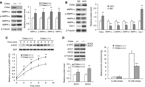
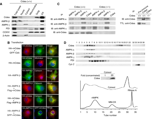
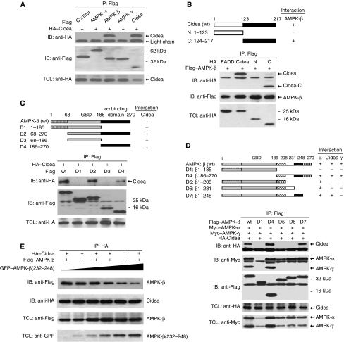
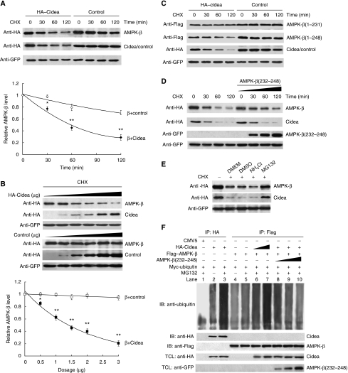
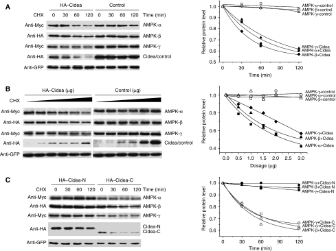
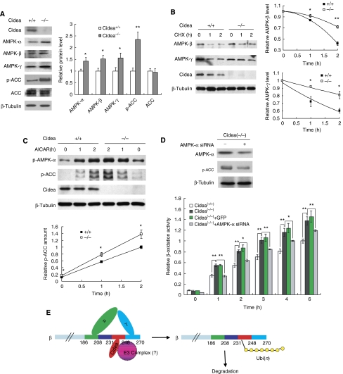
We previously showed that Cidea(-/-) mice are resistant to diet-induced obesity through the upregulation of energy expenditure. The AMP-activated protein kinase (AMPK), consisting of catalytic alpha subunit and regulatory subunits beta and gamma, has a pivotal function in energy homoeostasis. We show here that AMPK protein levels and enzymatic activity were significantly increased in the brown adipose tissue of Cidea(-/-) mice. We also found that Cidea is colocalized with AMPK in the endoplasmic reticulum and forms a complex with AMPK in vivo through specific interaction with the beta subunit of AMPK, but not with the alpha or gamma subunit. When co-expressed with Cidea, the stability of AMPK-beta subunit was dramatically reduced due to increased ubiquitination-mediated degradation, which depends on a physical interaction between Cidea and AMPK. Furthermore, AMPK stability and enzymatic activity were increased in Cidea(-/-) adipocytes differentiated from mouse embryonic fibroblasts or preadipocytes. Our data strongly suggest that AMPK can be regulated by Cidea-mediated ubiquitin-dependent proteosome degradation, and provide a molecular explanation for the increased energy expenditure and lean phenotype in Cidea-null mice.


Figures

Figure 1
Figure 1
Increased AMPK levels and enhanced AMPK activity in BAT of Cidea−/− mice. (A) Increased levels of AMPK-α, -β, -γ and phospho-AMPK-α in BAT of Cidea−/− mice. BAT was collected from 3-month-old wild-type and Cidea−/− male mice. β-Tubulin was served as a loading control. Similar experiments were carried out five times and the intensity of individual band in each western blot was quantified by TOTAL-LAB software (Nonlinear Dynamics, UK) and used for statistical analysis. The relative protein level in wild-type mice was designated as 1.0. **P<0.01. Similar quantitative and statistical analyses were conducted for all western blots shown in following figures. (B) BAT of high-fat diet (HFD)-treated mice had increased Cidea level and deceased AMPK levels compared with that of normal diet (ND)-fed animals. Similar experiments were conducted five times from five pairs of mice. The relative protein level of Cidea, Ucp1 and AMPK in mice fed with ND was designated as 1.0. (C) BAT of Cidea−/− mice showed enhanced AMPK activity. The upper panel was the time course of AMPK activity in BAT of wild-type and Cidea−/− mice. The lower panel was the quantitative analysis of the phospho-ACC western bolt bands from four independent experiments. The relative intensity of phospho-GST-ACC in wild-type mice at 8 min was designated as 1.0. (D) BAT of Cidea−/− mice showed increased endogenous ACC phosphorylation (left panel) and decreased ACC enzymatic activity (right panel). Four independent experiments were conducted using four pairs of mice (*P<0.05, **P<0.01, ***P<0.001).
Figure 2
Figure 2
Cidea and AMPK are localized to ER and form a complex in vivo. (A) Cidea and AMPK are present in ER-enriched fraction. Protein disulphide isomerase (PDI), β-actin and COXIV were used as specific markers for ER, cytosol and mitochondria, respectively. (B) Cidea is localized to ER, and AMPK-β is colocalized with Cidea when co-expressed. HA-tagged mouse Cidea (HA–mCidea), HA–AMPK-β and endogenous Calnexin were visualized by immunofluorescence. Fields shown (magnification, × 600) were visualized under fluorescence microscope at appropriate wavelengths for GFP (green), rhodamine (red) and Hoechst (blue), and the images were overlaid (merge, yellow). Plasmid DNAs used for transfection and immunostaining (transfection) were listed on the left side of each fluorescent staining image. (C) Cidea interacts with AMPK in vivo. Left panel, IP: immunoprecipitation using antibodies against AMPK-α, -β, -γ or Cidea, respectively. IB: immunoblot to detect AMPK-α, -β, -γ or Cidea in the immunoprecipitated products. Right panel, immunoprecipitation of Cidea by antibody against Cidea but not pre-immune serum. TTL: total tissue lysate. (D) Co-fractionation of Cidea and AMPK in cytosolic fraction of BAT. Bacterially expressed rat AMPK (rAMPK) complex (130 kDa), recombinant muscle-type creatine kinase (MM-CK; 86 kDa) and RNase A (14 kDa) were used as loading controls for gel filtration analysis. PDI (55 kDa) and cytochrome C (cyt C; 13 kDa) were used as controls for western blot analysis. Proteins from each eluted fraction were frozen-dried to concentrate approximately 20-fold and used for western blot analysis (insert of lower panel). Numbers on western blot correspond to the eluted fraction numbers from gel filtration chromatography.
Figure 3
Figure 3
Specific interaction between Cidea and AMPK-β. (A) Cidea interacts with AMPK-β but not -α and -γ. HA–Cidea was co-transfected with Flag–AMPK-α, -β or -γ into HEK 293T cells. AMPK-α, -β, -γ or Cidea were immunoprecipitated (IP) by antibody against Flag. Immunoprecipitated products were detected by immunoblotting (IB) against HA and flag antibodies. HA–Cidea was used as a positive control,whereas Flag–JNK1 as a negative control. TCL, total cell lysate. (B) Mapping of the interface on Cidea that mediates the interaction between Cidea and AMPK-β. The upper panel shows a schematic diagram depicting different Cidea truncations. Flag–AMPK-β was co-transfected with HA-tagged full-length Cidea or its truncations (N and C). AMPK-β was immunoprecipitated (IP) using antibody against Flag and the co-precipitated products were detected by immunoblotting (IB) using antibody against HA. Fas-associated protein with death domain (FADD) was used as a negative control. (C, D) Identification of regions of AMPK-β crucial for mediating interactions with Cidea, AMPK-α and -γ. Schematic diagram (upper panels) depicted different AMPK-β truncations. Flag–AMPK-β deletions and HA-tagged full-length Cidea were co-transfected. Immunoprecipitation (IP) was carried out using antibody against Flag and the co-immunoprecipitated products were detected by immunoblotting (IB) using antibody against HA, Flag or Myc. (E) Truncated AMPK-β containing aa 232–248 blocked the interaction between Cidea and AMPK-β. Increasing amounts of GFP-AMPK-β(232–248) were co-transfected with HA–Cidea and Flag–AMPK-β. HA–Cidea was immunoprecipitated (IP) and their co-precipitated products were detected by immunoblotting (IB) using antibodies against HA or Flag. TCL, total cell lysate.
Figure 4
Figure 4
Cidea accelerates the degradation of AMPK-β. (A) AMPK-β protein is degraded rapidly in the presence of Cidea. HA–FADD was used as a negative control. HA–AMPK-β (1.0 μg) was co-transfected with 1.0 μg of HA–Cidea or HA–FADD. GFP was a transfection control. Cycloheximide (CHX) was added into the medium and cells were harvested at 0, 30, 60 and 120 min after CHX treatment. The AMPK-β level before CHX treatment was designated as 1 (*P<0.05, **P<0.01). (B) Enhanced AMPK-β degradation in the presence of increasing amounts of Cidea. HA–AMPK-β was co-transfected with increasing amounts of HA–Cidea or HA–FADD. Cells were treated with CHX for 2 h before harvesting (*P<0.05, **P<0.01). (C) Cidea-mediated AMPK-β degradation is dependent on their direct interaction. Flag–AMPK-β(1–231) or AMPK-β(1–248) was co-transfected with HA–Cidea or FADD (*P<0.05, **P<0.01). (D) AMPK-β(232–248) attenuates Cidea-mediated AMPK-β degradation. Increasing amounts of GFP–AMPK-β(232–248) were co-transfected with HA–Cidea and HA–AMPK-β. (E) Cidea-mediated AMPK-β degradation is dependent on the proteosomal activity. MG132 (10 μM), a proteosome-specific inhibitor; NH4Cl (10 mM), a lysosomal protease inhibitor. DMEM containing DMSO was used as a control. (F) Cidea promotes AMPK-β ubiquitination. Myc-ubiquitin (2.0 μg), 1.0 μg HA–Cidea and Flag–AMPK-β were single or co-transfected. MG132 was added to a final concentration of 10 μM for 2 h. Cidea and AMPK-β were immunoprecipitated using antibodies against HA or Flag, respectively. SDS (0.5%) was added to the immunoprecipitation buffer to disrupt the interaction between Cidea and AMPK-β. CMV5 vector was transfected as a negative control (lane 1). Cidea can be ubiquitinated and MG132 treatment resulted in higher accumulation of ubiquitinated Cidea (lanes 2 and 3, respectively); levels of AMPK-β ubiquitination in the absence or presence of MG132 (lanes 4 and 5). AMPK-β ubiquitination is enhanced in the presence of increasing amounts of Cidea (1.0 and 2.0 μg for lanes 6 and 7, respectively). The attenuation of AMPK-β ubiquitination is seen in the presence of GFP–AMPK-β(232–248) that competes for Cidea (from 1.0 to 8.0 μg, lanes 8–10).
Figure 5
Figure 5
Cidea promotes the degradation of AMPK complex. (A) Accelerated degradation of AMPK complex when co-expressed with Cidea. Each AMPK subunit (0.1 μg) (Myc–AMPK-α, HA–AMPK-β and Myc–AMPK-γ) and 1.0 μg of HA–Cidea or HA–FADD were used for co-transfection. Statistical analysis (right panel, P<0.001) was evaluated from five independent experiments. (B) Degradation of AMPK complex is dependent on the amount of Cidea. Increasing amounts of Cidea (from 0 to 2 μg) or FADD were co-transfected with AMPK complex. Experiments were repeated five times and showed significant difference (P<0.001). (C) C-Terminal region of Cidea (Cidea-C) can promote AMPK complex degradation. Cidea-N or Cidea-C (1.0 μg) was co-transfected with AMPK subunits. Experiment was repeated four times and showed significant difference, P<0.01.
Figure 6
Figure 6
Cidea−/− adipocytes showed increased AMPK protein levels, AMPK stability, activity and fatty acid oxidation rate. (A) Cidea−/− adipocytes had increased levels of AMPK-α, -β -γ and ACC phosphorylation. MEFs isolated from wild-type and Cidea−/− mouse embryos were induced to differentiation into a brown adipocyte like cells (M&M). Cells were collected after 8 days of differentiation and used for western blot analysis. Protein levels were normalized by β-tubulin. The right panel shows the quantitative analysis of the western blot bands. The relative protein level of each AMPK subunit, total ACC and p-ACC in wild-type adipocytes was designated as 1.0. Statistical P-value was calculated from three similar sets of experiments (*P<0.05, **P<0.01). (B) Increased AMPK stability in Cidea−/− adipocytes. MEFs 8 days after differentiation were treated with CHX (500 μg/ml) for 1 or 2 h. The relative protein level before CHX treatment (0 hour) was designated as 1.0. *P<0.05. (C) Increased basal and AICAR-induced AMPK phosphorylation and activity in Cidea−/− adipocytes. AICAR (2 mM) was added into differentiated cells for 1 or 2 h. Levels of endogenous phosphorylated AMPK-α, ACC1 and ACC2 were used to evaluate AMPK activity. The lower panel shows the quantitative analysis of the p-ACC western blot bands. Levels of p-ACC in wild-type adipocytes after 2 h of AICAR treatment was designated as 1.0. (D) Cidea−/− adipocytes showed increased fatty acid β-oxidation, which can be attenuated by siRNA against AMPK-α. The upper panel shows the efficiency of lentiviral-mediated AMPK-α knockdown by siRNA (Cidea(−/−)+AMPK-α siRNA) in Cidea−/− adipocytes. The fatty acid β-oxidation rate of wild-type adipocytes at 6 h was designated as 1.0. The statistical results came from four replicates (*P<0.05, **P<0.01). Lentivirus containing GFP (Cidea(−/−)+GFP) was severed as a negative control. (E) The proposed model for AMPK-β degradation. Ubi(n): polyubiquitination.

References

    1. Baba M, Hong SB, Sharma N, Warren MB, Nickerson ML, Iwamatsu A, Esposito D, Gillette WK, Hopkins RF III, Hartley JL, Furihata M, Oishi S, Zhen W, Burke TR Jr, Linehan WM, Schmidt LS, Zbar B (2006) Folliculin encoded by the BHD gene interacts with a binding protein, FNIP1, and AMPK, and is involved in AMPK and mTOR signaling. Proc Natl Acad Sci USA 103: 15552–15557 - PMC - PubMed
    1. Banerjee RR, Rangwala SM, Shapiro JS, Rich AS, Rhoades B, Qi Y, Wang J, Rajala MW, Pocai A, Scherer PE, Steppan CM, Ahima RS, Obici S, Rossetti L, Lazar MA (2004) Regulation of fasted blood glucose by resistin. Science 303: 1195–1198 - PubMed
    1. Baur JA, Pearson KJ, Price NL, Jamieson HA, Lerin C, Kalra A, Prabhu VV, Allard JS, Lopez-Lluch G, Lewis K, Pistell PJ, Poosala S, Becker KG, Boss O, Gwinn D, Wang M, Ramaswamy S, Fishbein KW, Spencer RG, Lakatta EG et al. (2006) Resveratrol improves health and survival of mice on a high-calorie diet. Nature 444: 337–342 - PMC - PubMed
    1. Chan SC, Lin SC, Li P (2007) Regulation of Cidea protein stability by the ubiquitin-mediated proteasomal degradation pathway. Biochem J 408: 259–266 - PMC - PubMed
    1. Chen Z, Guo K, Toh SY, Zhou Z, Li P (2000) Mitochondria localization and dimerization are required for CIDE-B to induce apoptosis. J Biol Chem 275: 22619–22622 - PubMed

Publication types