. 2008 Oct;15(20):1384-96.
doi: 10.1038/gt.2008.87. Epub 2008 May 15.

Visualization of targeted transduction by engineered lentiviral vectors

Affiliations

Visualization of targeted transduction by engineered lentiviral vectors

K-I Joo et al. Gene Ther. 2008 Oct.

Abstract

We have reported a method to target lentiviral vectors to specific cell types. This method requires the incorporation of two distinct molecules on the viral vector surface: one is an antibody that renders the targeting specificity for the engineered vector, and the other is a fusogenic protein that allows the engineered vector to enter the target cell. However, the molecular mechanism that controls the targeted infection needs to be defined. In this report, we tracked the individual lentiviral particles by labeling the virus with the GFP-Vpr fusion protein. We were able to visualize the surface-displayed proteins on a single virion as well as antibody-directed targeting to a desired cell type. We also demonstrated the dynamics of virus fusion with endosomes and monitored endosome-associated transport of viruses in target cells. Our results suggest that the fusion between the engineered lentivirus and endosomes takes place at the early endosome level, and that the release of the viral core into the cytosol at the completion of the virus-endosome fusion is correlated with the endosome maturation process. This imaging study sheds some light on the infection mechanism of the engineered lentivirus and can be beneficial to the design of more efficient gene delivery vectors.


Figures
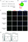
Figure 1
Figure 1
Co-incorporation of antibody and fusogenic protein on the single lentivirus particle. (A) The schematic representation of the labeling (GFP-Vpr) and viral (FUW and FUGW) constructs. CMV: cytomegalovirus immediate-early gene promoter; GFP: green fluorescence protein; Vpr: viral protein R; Ubi: human ubiqutin-C promoter; WPRE: woodchuck hepatitis virus posttranscriptional regulatory element. (B) The schematic representation of the virus-staining method for visualizing individual viruses. Two antibodies were used to detect the presence of αCD20 and fusogenic molecule (SINmu). (C) Colocalization of GFP-Vpr-labeled viral particles with the αCD20 antibody (red) and fusogenic molecule SINmu (blue). GFP-Vpr-labeled viruses psuedotyped by either both αCD20 and SINmu, or αCD20 antibody only, SINmu only, or VSVG protein were strained with anti-human IgG and anti-HA tag antibodies against αCD20 and SINmu. Overlapping green, red, and blue signals appears as white in a merged image. Scale bar represents 2 μm. (D) 293T/CD20 cells (2 × 10⁵) were transduced with 2 ml of fresh unconcentrated FUGW/αCD20+SINmu virus (∼ 1 × 10⁶ TU/ml) with or without GFP-Vpr labeling. The resulting GFP expression was analyzed by FACS. Solid line, analysis of the infected 293T/CD20; Shaded area, analysis of the non-infected 293T/CD20 (as a control).
Figure 2
Figure 2
Detection of virus-endosome fusion at different time points. (A) Schematic representation of the visualization assay for virus-endosome fusion by fluorescence dequenching. (B) The time course study of virus penetration (FUGW/VSVG and FUGW/αCD20+SINmu) by using bafilomycin A1 (see Materials and Methods) (C) GFP-Vpr-labeled VSVG or both αCD20 and SINmu displaying viruses (green) were labeled with DiD (red) for 1 h at room temperature. Double-labeled viruses were incubated with 293T/CD20 cells at 37°C for 0, 10, 30, or 60 min, fixed and imaged. The boxed regions are magnified and shown in separated panels below. Yellow particles indicate viral particles fused to endosomes. Scale bar represents 5 μm. (D & E) Tracking of endosomal fusion of individual viruses. (D) Selected images obtained from a time series study starting 30 min after incubation. The arrow indicates the viral particle monitored in the live cell. The GFP-Vpr+ (green) signal and GFP-Vpr+DiD+ (yellow as the merged color of green and red) signals mark the viral particle before and after endosomal fusion, respectively. Scale bar represents 2 μm. (E) Kinetics of the fluorescence intensity of the GFP-Vpr (green) and DiD (red) signals of the virion. The fluorescence intensity was measured within the regions of interests around viral particle using the software package for the Zeiss LSM 510.
Figure 3
Figure 3
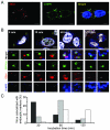
Internalization and transport of viruses through endosomes. (A) Distribution of endocytic compartments in living cells. Early and late endosomes were detected by EEA1 and CI-MPR, respectively. 293T/CD20 cells were immunostained with EEA1 (red) and CI-MPR (green) and counterstained with DAPI (blue). Arrows indicate individual endosomes positive for both early and late endosomal markers. (B) GFP-Vpr-labeled viruses (FUW-GFPVpr/αCD20+SINmu) were incubated with 293T/CD20 cells (MOI ∼30) at 37°C for various time points of 0, 30, 60, or 120 min. Then these cells were fixed, permeabilized, and immunostained with EEA1 (red) and CI-MPR (blue) and counterstained with DAPI (white). The boxed regions are magnified and shown in separated panels below. The bottom panels show the localization of GFP-Vpr-labeled viral particles in the two different endosomal stages. Scale bar represents 5 μm. (C) Quantification of GFP-Vpr-labeled lentiviruses colocalized with EEA1+ (black), CI-MPR+ (white), or EEA1+CI-MPR+ (gray) endosomes at different incubation times. The result shown is the collective data from three experiments.
Figure 4
Figure 4
Microtubule-associated transport of viruses. (A) Colocalization of GFP-Vpr-labeled lentiviruses with microtubule networks 1 h after infection. Microtubules were immunostained with the monoclonal antibody to α-tubulin (red). The boxed region is enlarged in right panel. Arrows indicate viruses on the microtubules. (B) Microtubule staining in a nocodazole-treated cell. Cells were preincubated with nocodazole at 37°C for 30 min to disrupt microtubules, then immunostained with the monoclonal antibody to α-tubulin (red). (C) The fixed image of GFP-Vpr/DiD-labeled viral particles in a nocodazole-treated cell (see Materials and Methods). Arrows indicate the viral particles fused to endosomes. (D) Localization of GFP-Vpr-labeled viral particles (FUW-GFPVpr/αCD20+SINmu) with the two endosomal markers after 60 min of incubation in a nocodazole pre-treated cell. (E) Microtubule staining of siRNA-treated cell. 293T/CD20 cells were transfected with α-tubulin-specific siRNA- or control siRNA. After 72 h, transfected cells were seeded and immunostained with anti- α-tubulin antibody (green). (F) The virus fusion for siRNA-treated cells. α-tubulin siRNA or control siRNA-treated cells were incubated with GFP-Vpr/DiD-labeled viruses at 37°C for 60 min and then fixed. Yellow particles denoted by arrows indicate the virus particles fused to endosomes. (G) Localization of GFP-Vpr-labeled viral particles with the two endosomal markers after 60 min of incubation with α-tubulin siRNA-treated cells. The boxed regions are enlarged and shown in separated panels. Scale bar represents 5 μm.
Figure 5
Figure 5
The effects of inhibitory drugs or siRNA treatment on viral fusion, infection, and endosome maturation. (A) GFP-Vpr/DiD-labeled viruses were incubated with drug- or siRNA-treated cells at 37°C for 60 min, and then fixed. The viral particles with the fusion signal (black) or without the fusion signal (gray) were quantified. The viral particles both GFP-Vpr+ and DiD+ were considered to be fused with endosomes, while particles that were only GFP-Vpr+ were considered to be unfused virus. For quantification, 60 viral particles were examined for no drug-treatment, 64 particles for nocodazole treatment, 72 particles for α-tubulin siRNA treatment, and 68 particles for cyto-D treatment. The results were collected from three independent experiments. (B) The role of microtubules and actin filaments in the virus infection. 293T/CD20 cells which were preincubated with nocodazole or cytochalasin-D (cyto-D) were transduced with 2 ml of fresh unconcentrated FUGW/αCD20+SINmu virus. The resulting GFP expression was analyzed by FACS. (C) Quantification of GFP-Vpr-labeled viruses colocalized with EEA1+ (black), CI-MPR+ (white), or both EEA1+ and CI-MPR+ (gray) endosomes at 60 min of incubation in drug- or siRNA-treated cells. (D) The effect of α-tubulin knockdown on virus infection. Control siRNA or α-tubulin siRNA transfected cells were transduced with 2 ml of fresh unconcentrated FUGW/αCD20+SINmu virus. The percentage of GFP+ cells was analyzed by FACS.
Figure 6
Figure 6
Time series images of the viral core release from an endosome starting 60 min after incubation. GFP-Vpr/DiD-labeled viruses (FUW-GFPVpr/αCD20+SINmu) were incubated with 293T/CD20 cells at 37°C for 60 min to initiate virus fusion, and then time-series images were obtained at every ∼10 seconds over a time period of 20 min. Scale bar represents 2 μm.

Similar articles

Cited by

References

    1. Daly G, Chernajovsky Y. Recent developments in retroviral-mediated gene transduction. Mol. Ther. 2000;2:423–434. - PubMed
    1. Mountain A. Gene therapy: the first decade. Trends Biotechnol. 2000;18:119–128. - PubMed
    1. Somia N, Verma IM. Gene therapy: Trials and tribulations. Nat. Rev. Genet. 2000;1:91–99. - PubMed
    1. Verma IM, Somia N. Gene therapy-promises, problems and prospects. Nature. 1997;389:239–242. - PubMed
    1. Bukrinsky MI, Sharova N, Dempsey MP, Stanwick TL, Bukrinskaya AG, Haggerty S, et al. Active nuclear import of human immunodeficiency virus type 1 preintegration complexes. Proc. Natl. Acad. Sci. USA. 1992;89:6580–6584. - PMC - PubMed

Publication types

MeSH terms