Comparative Study
. 1991 Mar 8;64(5):1007-15.
doi: 10.1016/0092-8674(91)90324-r.

Liquid-crystalline, phage-like packing of encapsidated DNA in herpes simplex virus

Affiliations
Comparative Study

Liquid-crystalline, phage-like packing of encapsidated DNA in herpes simplex virus

F P Booy et al. Cell. .

Abstract

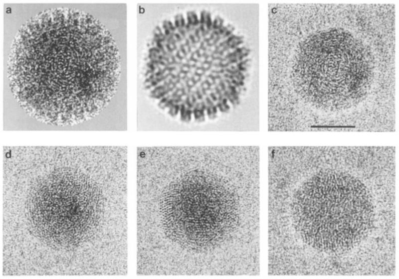
The organization of DNA within the HSV-1 capsid has been determined by cryoelectron microscopy and image reconstruction. Purified C-capsids, which are fully packaged, were compared with A-capsids, which are empty. Unlike A-capsids, C-capsids show fine striations and punctate arrays with a spacing of approximately 2.6 nm. The packaged DNA forms a uniformly dense ball, extending radially as far as the inner surface of the icosahedral (T = 16) capsid shell, whose structure is essentially identical in A-capsids and C-capsids. Thus we find no evidence for the inner T = 4 shell previously reported by Schrag et al. to be present in C-capsids. Encapsidated HSV-1 DNA closely resembles that previously visualized in bacteriophages T4 and lambda, thus supporting the idea of a close parallelism between the respective assembly pathways of a major family of animal viruses (the herpesviruses) and a major family of bacterial viruses.


Figures

Figure 1
Figure 1. Cryoelectron Micrographs of purified Preparatopms of A-Capsids and C-Capsids of HSV-1
(a) A-capsids. (b) C-capsids. Viewed at higher magnification (c), C-capsids show a distinctive “fingerprint” motif of punctate arrays or finely spaced, curvilinear striations. Bar = 100 nm
Figure 2
Figure 2. Stereo Pairs of the Outer Surface of a Three-Dimensional Reconstruction of C-Capsids of HSV-I
The particles are viewed along a 5-fold axis of symmetry (upper panels) and a 3-fold axis of symmetry (lower panels).
Figure 3
Figure 3. Transverse Central Sections Taken from the Three-Dimensional Density Maps of C-Capsids and A-Capsids of HSV-1, Reconstructed from Cryoelectron Micrographs
(a) C-capsid. (b) A-capsid. The particles are oriented as if viewed along a2-fold symmetry axis. The respective outer shell structures closely resemble each other. In the empty A-capsids, the density inside is typically at the same level as the solvent outside. In C-capsids, the density inside is much higher than background but lower than in the shell on account of averaging (see Discussion). In both cases, although the internal structure (or solvent) is not icosahedrally symmetric, the sections show, with low contrast, internal features whose symmetry was imposed by the icosahedral reconstruction procedure. Bar = 50 nm.
Figure 4
Figure 4. Computer-Filtered images of Cores of C-Capsids of HSV-I
For a typical particle (a), panel (b) represents the contribution of the icosahedral shell to this image, calculated by reprojecting its three-dimensional reconstruction into this viewing geometry. The shell contribution is then digitally subtracted from the original to expose the core (c). All such filtered images show the characteristic “fingerprint” motif of packaged HSV-1 DNA. Panels (d)–(f) show three more examples. The vortex-like aspect of fingerprints is illustrated in (c) and (d), punctate arrays in (d) and (e), and linear striations as well as punctate formations in (f). Bar = 50 nm.
Figure 5
Figure 5. Powder Pattern Diffractograms Calculated from Cryoelectron Micrographs of HSV-1 Capsids
(a) Composite pattern from 16 C-capsids, showing a system of concentric rings. Like the individual patterns (not shown), the composite pat-Iern is azimuthally symmetric, indicating a stigmatic micrograph. The intensity falls to zero at a spatial frequency of about (2.3 nm)−1, which is the effective resolution limit of these data, there being only a very faint secondary lobe of contrast transfer. The composite patterns were azimuthally averaged to reduce the noise, and the resulting C-capsid pattern (c) is compared with the A-capsid pattern (b). The particles used in this analysis were all present in the same untilted micrograph, so that this comparison is not biased by any differences in defocus, magnification, etc. The difference spectrum (d) represents primarily the contribution of the packaged DNA. Its peak, at (2.6 nm)−1, marks the average interduplex spacing (arrow).
Figurd 6
Figurd 6. Protein Compositions of A-Capsids and C-Capsids
SDS-polyacrylamide gel electrophoretogram, stained with Coomassie blue, comparing the protein compositions of A-capsids and C-capsids of HSV-1. The major components are indicated. The two protein compositions are essentially identical.

References

    1. Baker TS, Drak J, Bina M. Reconstruction of the three-dimensional structure of simian virus 40 and visualization of the chromatin core. Proc. Natl. Acad. Sci. USA. 1988;85:422–426. - PMC - PubMed
    1. Baker TS, Drak J, Bina M. The capsid of small papova viruses contains 72 pentameric capsomeres: direct evidence from cryo-electron microscopy of simian virus 40. Biophys. J. 1989;55:243–253. - PMC - PubMed
    1. Baker TS, Newcomb WW, Booy FP, Brown JC, Steven AC. Three-dimensional structures of maturable and abortive capsidsofequine herpesvirus 1 from cryoelectron microscopy. J. Virol. 1990;64:563–573. - PMC - PubMed
    1. Black LW, Newcomb WW, Boring JW, Brown JC. Ion etching of bacteriophage T4: support for a spiral-fold model of packaged DNA. Proc. Natl. Acad. Sci. USA. 1985;82:7960–7964. - PMC - PubMed
    1. Black LW. DNA packaging in dsDNA bacteriophages. Annu. Rev. Microbial. 1989;43:267–292. - PubMed

Publication types