Review
. 2008 Aug;85(1):11-9.
doi: 10.1016/j.yexmp.2008.03.007. Epub 2008 Apr 8.

Origins and virulence mechanisms of uropathogenic Escherichia coli

Affiliations
Review

Origins and virulence mechanisms of uropathogenic Escherichia coli

Travis J Wiles et al. Exp Mol Pathol. 2008 Aug.

Abstract

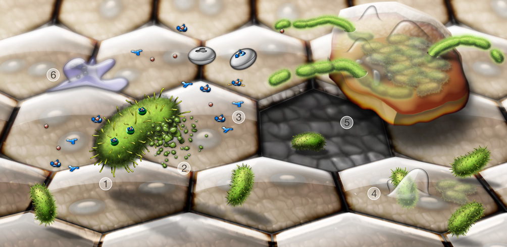
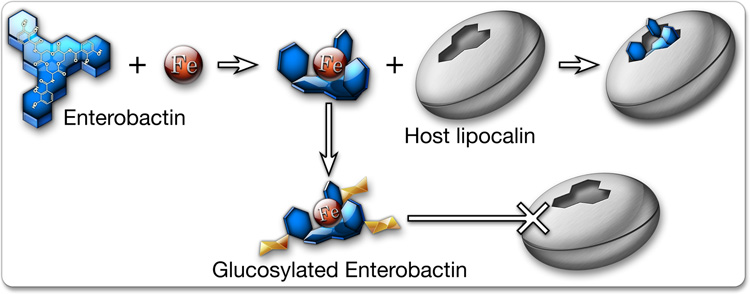
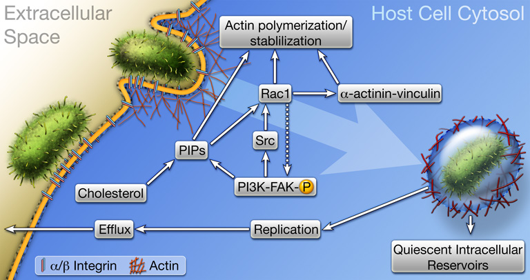
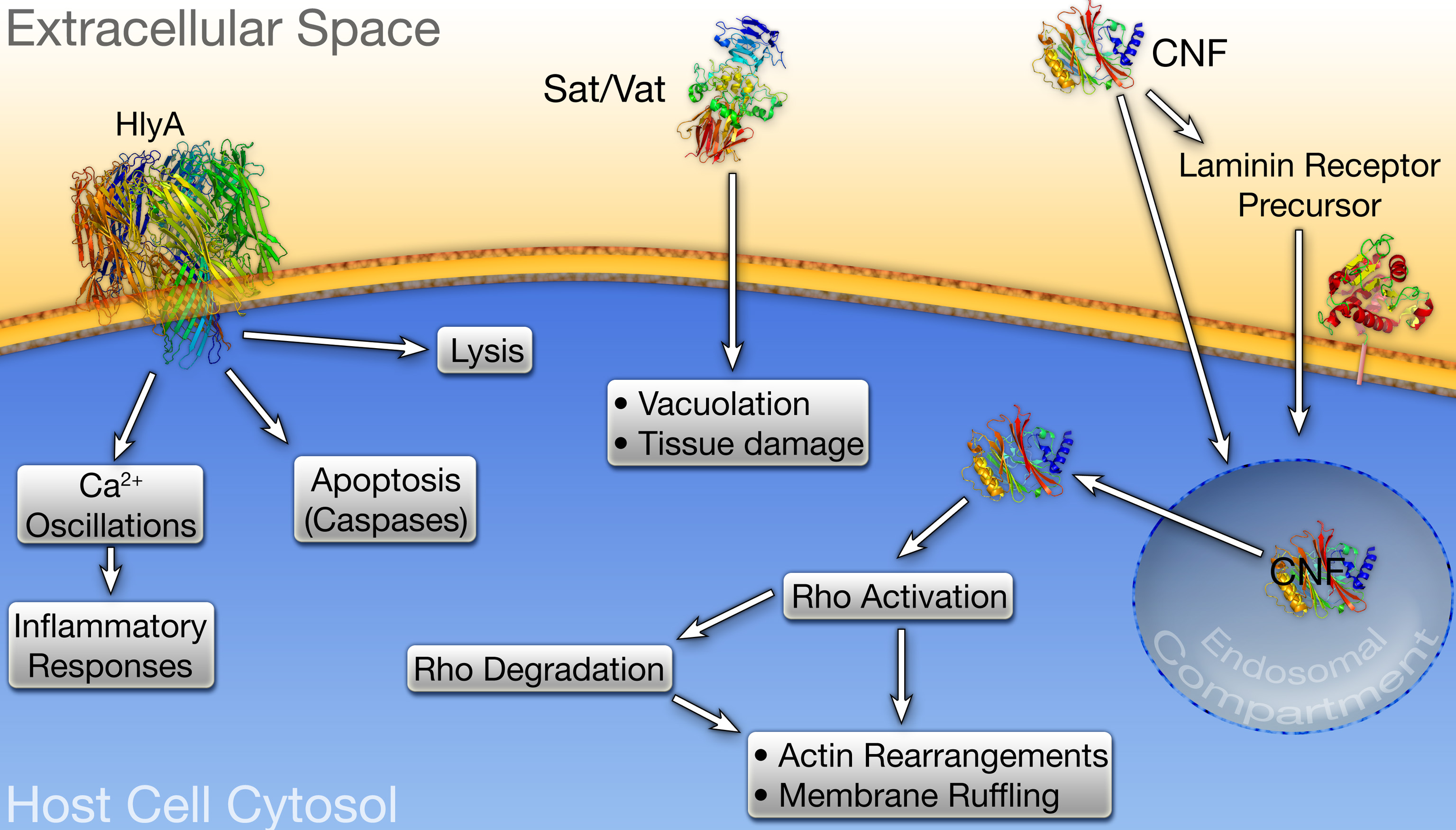
Strains of uropathogenic E. coli (UPEC) are the primary cause of urinary tract infections, including both cystitis and pyelonephritis. These bacteria have evolved a multitude of virulence factors and strategies that facilitate bacterial growth and persistence within the adverse settings of the host urinary tract. Expression of adhesive organelles like type 1 and P pili allow UPEC to bind and invade host cells and tissues within the urinary tract while expression of iron-chelating factors (siderophores) enable UPEC to pilfer host iron stores. Deployment of an array of toxins, including hemolysin and cytotoxic necrotizing factor 1, provide UPEC with the means to inflict extensive tissue damage, facilitating bacterial dissemination as well as releasing host nutrients and disabling immune effector cells. These toxins also have the capacity to modulate, in more subtle ways, host signaling pathways affecting myriad processes, including inflammatory responses, host cell survival, and cytoskeletal dynamics. Here, we discuss the mechanisms by which these and other virulence factors promote UPEC survival and growth within the urinary tract. Comparisons are also made between UPEC and other strains of extraintestinal pathogenic E. coli that, although closely related to UPEC, are distinct in their abilities to colonize the host and cause disease.


Figures

Figure 1
Figure 1
Dynamic interplay between invading UPEC and the host during a UTI. Shown are key events taking place during bladder infection by UPEC. Type 1 pili-expressing UPEC (1, green) secrete toxins and other virulence factors, alone or in association with outer membrane vesicles (2). Siderophores like enterobactin and salmochelin (3, blue structures) released by UPEC scavenge iron, in competition with host iron chelating molecules and lipocalin 2 (white discs). Type 1 pili mediate bacterial attachment to and invasion of the bladder epithelial cells (4). Large terminally differentiated superficial epithelial cells, which are often binucleate and have distinctive hexagonal or pentagonal shapes, line the lumenal surface of the bladder and are the primary targets of UPEC invasion. UPEC can rapidly multiply within the superficial cells, forming large biofilm-like communities. Exfoliation of infected bladder cells facilitates bacterial clearance from the host, but leaves the smaller underlying immature cells more susceptible to infection (5). The release, or efflux, of UPEC from infected host cells before they complete exfoliation likely promotes bacterial dissemination and persistence within the urinary tract. During efflux, UPEC often become filamentous, probably due in part to mounting stress arising from increased activation of host defenses. These include the influx of neutrophils (6), as well as the generation of reactive oxygen and nitrogen species and antimicrobial peptides.
Figure 2
Figure 2
Iron acquisition. Both host and pathogen compete for iron and have evolved multiple strategies to outdo the other. The bacterial siderophore enterobactin sequesters iron with high affinity, while host lipochalin 2 binds enterobactin and prevents its uptake by UPEC. Bacteria carrying the iroA gene cluster can modify enterobactin by glucosylation, creating salmochelin, which effectively binds iron but is not recognized by lipochalin 2.
Figure 3
Figure 3
Host cell invasion by UPEC. The FimH adhesin localized at the distal tips of type 1 pili engages α3β1 integrin receptors, and possibly other receptors, which likely cluster within cholesterol-rich lipid rafts. Receptor binding triggers signaling cascades involving FAK, Src, PI 3-kinase, Rho GTPases like Rac1, phosphoinositides (PIPs), and transient complex formation between the cytoskeleton stabilizing and scaffolding proteins α-actinin and vinculin. These events stimulate actin rearrangements, causing the host plasma membrane to zipper around and envelope bound bacteria. Once internalized, UPEC can be trafficked to late endosome-like compartments that are often localized within a meshwork of actin filaments. Bacteria exist quiescently within these actin-bound compartments and may serve as reservoirs for recurrent UTIs. Liberation of UPEC into the host cytosol stimulates rapid bacterial growth and the formation of intracellular biofilm-like communities.
Figure 4
Figure 4
UPEC-associated toxins. α-hemolysin (HlyA), Vat, Sat, and cytotoxic necrotizing factor 1 (CNF1) are encoded by many UPEC isolates. (left) At high concentrations, HlyA inserts into the membrane of target host cells and promotes their lysis, whereas at sublytic concentrations HlyA can modulate signaling cascades affecting host cell survival and inflammatory responses. (middle) Intoxication of host cells by Vat or Sat induces vacuolation and other cytopathic effects, leading to host tissue damage. (right) Endocytosis of CNF1 occurs through the laminin receptor precursor, or via alternative delivery mechanisms in association with outer membrane vesicles. Conditions within the endosomal compartment stimulate translocation of the CNF1 catalytic domain into the host cytosol where it activates Rho GTPases, inducing their eventual degradation subsequent membrane ruffling.

References

    1. Allen PM, et al. Contribution of capsular polysaccharide and surface properties to virulence of Escherichia coli K1. Infect Immun. 1987;55:2662–2668. - PMC - PubMed
    1. Anderson GG, et al. Intracellular bacterial biofilm-like pods in urinary tract infections. Science. 2003;301:105–107. - PubMed
    1. Andrews SC, et al. Bacterial iron homeostasis. FEMS Microbiol Rev. 2003;27:215–237. - PubMed
    1. Bahrani-Mougeot FK, et al. Type 1 fimbriae and extracellular polysaccharides are preeminent uropathogenic Escherichia coli virulence determinants in the murine urinary tract. Mol Microbiol. 2002;45:1079–1093. - PubMed
    1. Balsalobre C, et al. Release of the type I secreted alpha-haemolysin via outer membrane vesicles from Escherichia coli. Mol Microbiol. 2006;59:99–112. - PubMed

Publication types

MeSH terms

Substances