Review
. 2008 Aug 7;149(1-3):15-25.
doi: 10.1016/j.regpep.2007.08.024. Epub 2008 Mar 25.

The vagus nerve, food intake and obesity

Affiliations
Review

The vagus nerve, food intake and obesity

Hans-Rudolf Berthoud. Regul Pept. .

Abstract

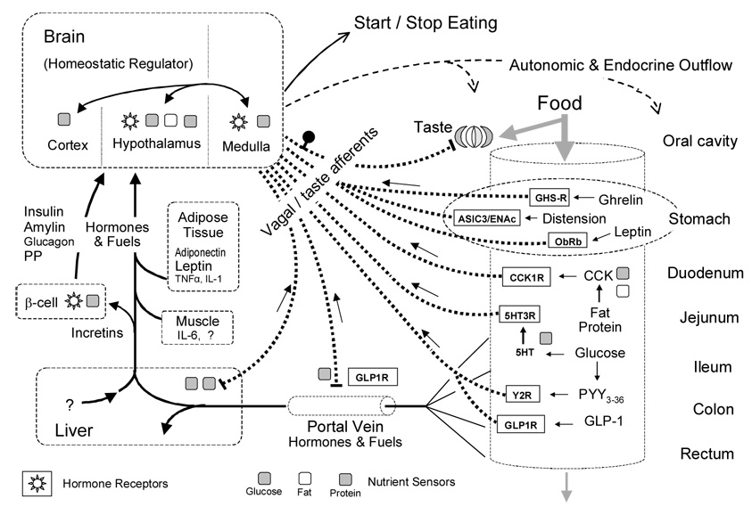
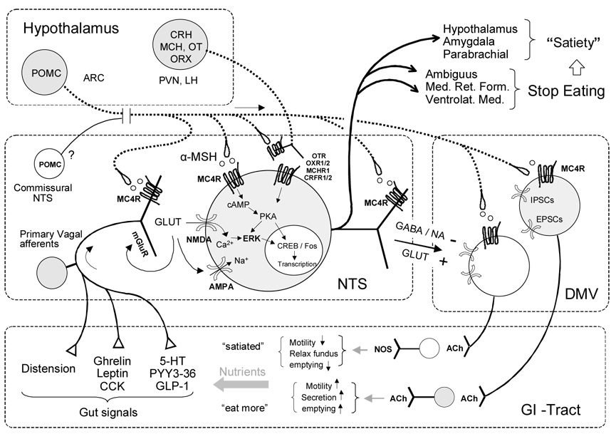
Food interacts with sensors all along the alimentary canal to provide the brain with information regarding its composition, energy content, and beneficial effect. Vagal afferents innervating the gastrointestinal tract, pancreas, and liver provide a rapid and discrete account of digestible food in the alimentary canal, as well as circulating and stored fuels, while vagal efferents, together with the sympathetic nervous system and hormonal mechanisms, codetermine the rate of nutrient absorption, partitioning, storage, and mobilization. Although vagal sensory mechanisms play a crucial role in the neural mechanism of satiation, there is little evidence suggesting a significant role in long-term energy homeostasis. However, increasing recognition of vagal involvement in the putative mechanisms making bariatric surgeries the most effective treatment for obesity should greatly stimulate future research to uncover the many details regarding the specific transduction mechanisms in the periphery and the inter- and intra-neuronal signaling cascades disseminating vagal information across the neuraxis.


Figures

Fig. 1
Fig. 1. Nutrient sensing in the alimentary canal and the control of food intake
Simplified schematic diagram showing the major pre- and postabsorptive transduction sites and mechanisms for the detection of ingested food and its macronutrient components. Nutrient information is sent to the brain through vagal and taste afferents (heavy dotted lines) or through the blood circulation (full lines). Specific receptors expressed by vagal afferent neurons are shown in rectangular boxes. Specific sensor mechanisms demonstrated for glucose, amino acids/proteins, and lipids/fatty acids are shown by gray, striped, and white squares, respectively.
Fig. 3
Fig. 3. Simplified schematic diagram showing the neural systems responsible for satiation and meal size control
Hypothetical neuron in the nucleus tractus solitarius (NTS) receives information from gastrointestinal mechano- and chemo-sensors through vagal afferents and projects back to the gut via vago-vagal reflexes through the dorsal motor nucleus (DMV). Other outputs (heavy lines) of certain NTS neurons are directed towards the medullary reticular formation (Med. Ret. Form.) and eventually to brainstem motor nuclei responsible for oromotor control necessary to start and stop eating, as well as to forebrain areas, responsible for sustained satiety. Descending modulatory projections from hypothalamic areas are also shown (heavy dotted lines).
Fig. 4
Fig. 4. Schematic diagram showing implications regarding vagal innervation and endocrine functions of gastric bypass surgery
The nutrient limb of the Rou-xen-Y gastric bypass consisting of the small gastric pouch and the anastomosed jejunum is shown on the right (shaded). The bilio-pancreatic limb including the large gastric remnant with attached duodenum and proximal jejunum is shown on the left. Note that the stomach remnant is depicted at a much reduced size for clarity. The ventral (anterior) and dorsal (posterior) vagal trunks and their branches are shown as solid and dotted lines, respectively (the severed ventral gastric branches are in light gray). The relative density and distribution of enteroendocrine cells secreting peptide hormones or transmitters are depicted by different symbols as indicated.

References

    1. Abell TL, Minocha A, Abidi N. Looking to the future: electrical stimulation for obesity. Am J Med Sci. 2006;331:226–232. - PubMed
    1. Alessi NE, Quinlan P, Khachaturian H. MSG effects on beta-endorphin and alpha-MSH in the hypothalamus and caudal medulla. Peptides. 1988;9:689–695. - PubMed
    1. Andersen DK, Elahi D, Brown JC, Tobin JD, Andres R. Oral glucose augmentation of insulin secretion. Interactions of gastric inhibitory polypeptide with ambient glucose and insulin levels. J Clin Invest. 1978;62:152–161. - PMC - PubMed
    1. Anini Y, Hansotia T, Brubaker PL. Muscarinic receptors control postprandial release of glucagons-like peptide-1: in vivo and in vitro studies in rats. Endocrinology. 2002;143:2420–2426. - PubMed
    1. Azzara AV, Sokolnicki JP, Schwartz GJ. Central melanocortin receptor agonist reduces spontaneous and scheduled meal size but does not augment duodenal preload-induced feeding inhibition. Physiol Behav. 2002;77:411–416. - PubMed