. 2008 Aug 15;283(33):22709-22.
doi: 10.1074/jbc.M801319200. Epub 2008 May 15.

Potential molecular mechanism for c-Src kinase-mediated regulation of intestinal cell migration

Affiliations

Potential molecular mechanism for c-Src kinase-mediated regulation of intestinal cell migration

Sijo Mathew et al. J Biol Chem. .

Abstract

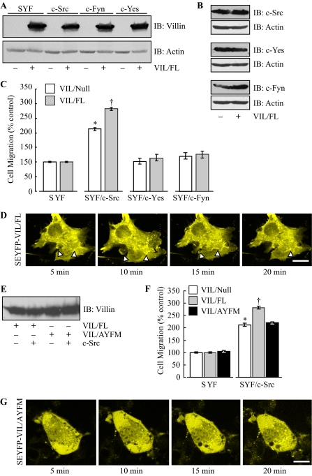
The ubiquitously expressed Src tyrosine kinases (c-Src, c-Yes, and c-Fyn) regulate intestinal cell growth and differentiation. Src activity is also elevated in the majority of malignant and premalignant tumors of the colon. The development of fibroblasts with the three ubiquitously expressed kinases deleted (SYF cells) has identified the role of Src proteins in the regulation of actin dynamics associated with increased cell migration and invasion. Despite this, unexpectedly nothing is known about the role of the individual Src kinases on intestinal cell cytoskeleton and/or cell migration. We have previously reported that villin, an epithelial cell-specific actin-modifying protein that regulates actin reorganization, cell morphology, cell migration, cell invasion, and apoptosis, is tyrosine-phosphorylated. In this report using the SYF cells reconstituted individually with c-Src, c-Yes, c-Fyn, and wild type or phosphorylation site mutants of villin, we demonstrate for the first time the absolute requirement for c-Src in villin-induced regulation of cell migration. The other major finding of our study is that contrary to previous reports, the nonreceptor tyrosine kinase, Jak3 (Janus kinase 3), does not regulate phosphorylation of villin or villin-induced cell migration and is, in fact, not expressed in intestinal epithelial cells. Further, we identify SHP-2 and PTP-PEST (protein-tyrosine phosphatase proline-, glutamate-, serine-, and threonine-rich sequence) as negative regulators of c-Src kinase and demonstrate a new function for these phosphatases in intestinal cell migration. Together, these data suggest that in colorectal carcinogenesis, elevation of c-Src or down-regulation of SHP-2 and/or PTP-PEST may promote cancer metastases and invasion by regulating villin-induced cell migration and cell invasion.


Figures

FIGURE 1.
FIGURE 1.
c-Src kinase is required for the intracellular localization of villin at the cell margin. A, Src tyrosine kinase activity was monitored in vitro by measuring phosphorylation of enolase. Phosphorylation of enolase was detected by Western analysis using phosphotyrosine antibody (PY-20). These data are representative of two experiments with similar results. B, recombinant villin protein was phosphorylated in vitro with Src kinases immunoprecipitated from SYF/c-Src, SYF/c-Yes, and SYF/c-Fyn cells, respectively. Immunoprecipitates in the absence of primary antibodies to c-Src, c-Yes, and c-Fyn were run as negative controls (from left, lanes 3). These data are representative of three other experiments with similar results. C, SYF, SYF/c-Src, SYF/c-Yes, and SYF/c-Fyn cells were transiently transfected with SEYFP-VIL/FL. In addition, SYF/c-Src cells were transiently transfected with villin mutant, SEYFP-VIL/ANFM. Bar, 10 μm. *, p < 0.01, n = 20. D, Caco-2 cells were infected with recombinant adenovirus to express either DN c-Src or Ad-EGFP (control). Bar, 10 μm. IB, immunoblot.
FIGURE 2.
FIGURE 2.
c-Src kinase is required for villin-induced increase in cell migration. A, cells were infected with recombinant adenovirus to express vector alone (-VIL/FL) or full-length villin protein (+VIL/FL). Western blots are representative of three other experiments with similar results. B, this Western blot shows comparable levels of c-Src, c-Yes, and c-Fyn in all three cell lines used for this study. C, cell migration was recorded in cells expressing recombinant adenoviral vector (VIL/Null) or full-length villin protein (VIL/FL). Values are means ± S.E. (n = 12). *, p < 0.01, statistically significant compared with control cells. †, p < 0.01, statistically significant compared with SYF/c-Src cells expressing vector alone. D, SYF/c-Src cells expressing SEYFP-VIL/FL were treated with EGF (100 ng/ml), and time-lapse images were recorded 5 min after the addition of EGF for a total of 20 min. Lamellipodia and membrane ruffles are marked with arrowheads. Bar, 10 μm. E, cells were infected with adenovirus to express vector (-VIL/FL), VIL/FL, or VIL/AYFM. F, cell migration rates were recorded in SYF and SYF/c-Src cells expressing either vector alone (VIL/Null), VIL/FL, or VIL/AYFM. Values are means ± S.E. *, p < 0.01, statistically significant compared with control cells. †, p < 0.01, statistically significant compared with SYF/c-Src cells infected with vector alone. G, SYF/c-Src cells expressing SEYFP-VIL/AYFM were treated with EGF (100 ng/ml), and time lapse images were recorded 5 min after the addition of EGF for a total of 20 min. Bar, 10 μm. IB, immunoblot.
FIGURE 3.
FIGURE 3.
c-Src kinase regulates villin-induced increase in cell invasion. Cell invasion was recorded in HeLa Tet-Off cells stably transfected with full-length human villin cultured in the presence (VIL/Null) or absence (VIL/FL) of doxycycline. Bar, 20 μm. In the presence of villin, there was a statistically significant increase in the number of cells that invade through the Matrigel (†, p < 0.001) compared with VIL/Null cells. Pretreatment of VIL/Null cells with the Src kinase inhibitor PP2 (100 nm) (A) or expression of dominant-negative c-Src kinase in VIL/Null cells (DN c-Src) (B) inhibited cell invasion (*, p < 0.05 compared with VIL/Null cells in the absence of PP2 and in VIL/Null cells infected with adenovirus to express Ad-EGFP, respectively). Pretreatment of VIL/FL cells with PP2 (A) or infection of VIL/FL cells with recombinant adenovirus to express DN c-Src (B) significantly inhibited cell invasion (#, p < 0.001 compared with VIL/FL cells in the absence of PP2 and compared with VIL/FL cells infected with adenovirus to express Ad-EGFP, respectively).
FIGURE 4.
FIGURE 4.
Jak3 does not regulate tyrosine phosphorylation of villin or villin-induced cell migration. A, SYF/Jak3 cells were infected with recombinant adenovirus to express either vector alone (-VIL/FL) or VIL/FL. Western blots are representative of three other experiments with similar results. B, the tyrosine kinase activity of transfected Jak3 was monitored by measuring phosphorylation in vitro of GST-γc protein. GST was used as a negative control. C, cell migration rates were recorded in SYF and SYF/Jak3 cells expressing recombinant adenoviral vector (VIL/Null) or VIL/FL. n = 12. D, Caco-2 cell migration was measured in the absence or presence of Jak3 inhibitor VI (54 nm). Cell migration was recorded in MDCK Tet-Off VIL/Null and VIL/FL cells in the absence or presence of Jak3 inhibitor (13 nm). *, p < 0.01, statistically significant compared with control cells. E, in vitro tyrosine phosphorylation of recombinant villin protein was measured using Jak3 and c-Src kinases, immunoprecipitated from SYF/Jak3 and SYF/c-Src cells, respectively. This Western blot is a representative of three other experiments with similar results. F, recombinant full-length villin was tyrosine-phosphorylated in vitro using recombinant c-Src kinase and recombinant Jak3 kinase. From the left, lane 3 shows no phosphorylation of recombinant villin by recombinant Jak3; lane 4 shows autophosphorylation of recombinant c-Src kinase; lane 5 shows tyrosine phosphorylation of recombinant villin (upper band) by recombinant c-Src kinase (lower band, autophosphorylated c-Src). This Western is a representative of three other experiments with similar results. G, expression of Jak3 in the intestinal cell lines, Caco-2 and HT29/19A, and in the renal epithelial cell line, MDCK. SYF/Jak3 cell lysates were used as a positive control. This is a representative of four experiments with similar results. H, Caco-2 and SYF/Jak3 cell lysates were used to immunoprecipitate (IP) Jak3. The arrow indicates the Jak3 protein identified in SYF/Jak3 cell lysates and immunoprecipitates but not in Caco-2 cells. This is a representative of two experiments with similar results. I, histological analysis of mouse ileum shows Jak3 staining in the submucosa, but none of the epithelial cells in the villi stained positive for Jak3 (a). A negative control stained in the absence of primary antibody is shown below (b). This is a representative histological specimen, and similar data were obtained in three other experiments. Bar, 100 μm. J, RT-PCR analysis shows that Jak3 mRNA expression is not detectable in HT29/19A or Caco-2 cells. Jurkat cells were used as a positive control. S15 transcript was amplified from the same template as a reference control. Jak3 genomic DNA contamination was ruled out by PCR amplification from the same RNA template. NC, negative control; reaction mixture containing all ingredients except RNA template. IB, immunoblot; IP, immunoprecipitation.
FIGURE 5.
FIGURE 5.
Syk2 does not regulate tyrosine phosphorylation of villin or villin-induced cell migration. A, SYF cells were infected with recombinant adenovirus to express either vector alone (-VIL/FL) or full-length human villin (+VIL/FL). Western blots are representative of three other experiments with similar results. B, cell migration rates were recorded in SYF and SYF/Syk2 cells expressing vector (VIL/Null) or VIL/FL. n = 12. C, in vitro phosphorylation of recombinant villin by Syk2. Phosphorylation of recombinant villin by c-Src was used as a positive control in this assay (from left, lane 3). This Western blot is a representative of three other experiments. IB, immunoblot.
FIGURE 6.
FIGURE 6.
Protein tyrosine phosphatases, SHP-2 and PTP-PEST, regulate villin-induced cell migration by regulating the catalytic inactivation of c-Src kinase. A, SYF/c-Src cells were infected with adenovirus to express vector alone (-VIL/FL) or full-length human villin (+VIL/FL) with either SHP-2 or PTP-PEST. Western blots are representative of three other experiments with similar results. B, SHP-2 and PTP-PEST catalytic activity is expressed in μmol/min. Analysis of cell lysates from three 6-well plates run in triplicates are shown. Data show a statistically significant (*, p < 0.001, n = 6) increase in phosphatase activity in cells infected with constitutively active SHP-2 and PTP-PEST compared with their respective control cells expressing vector alone. C, SYF/c-Src cells expressing SHP-2 or PTP-PEST transfected with SEYFP-VIL/FL. Bar, 10 μm. D, SYF cells were infected with recombinant adenovirus to express vector alone or VIL/FL, and cell migration rates were determined. E, cell migration rates were determined in SYF/c-Src cells infected with recombinant adenovirus to express vector alone (VIL/Null) or VIL/FL without or with SHP-2 or PTP-PEST. Values are means ± S.E. (n = 12). †, p < 0.01, statistically significant compared with control cells. *, p < 0.01, statistically significant compared with SYF/c-Src cells expressing full-length villin. F, cell migration rates were recorded in SYF and SYF/c-Src cells infected with adenovirus to express vector (VIL/Null) or VIL/FL without or with SHP-2 and PTP-PEST. Values are means ± S.E. (n = 12). *, p < 0.01, statistically significant compared with cells expressing vector alone; †, p < 0.01 statistically significant compared with SYF/c-Src cells expressing vector alone. G, cell migration was measured in Caco-2 cells overexpressing SHP-2 and/or PTP-PEST. Values are means ± S.E. (n = 12). *, p < 0.01, statistically significant compared with control cells. H (left), dephosphorylation of recombinant tyrosine-phosphorylated villin by PTP-PEST or SHP-2. Alkaline phosphatase (2.5 units) was used as a positive control (from left, lanes 5 and 6). These Western blots are representative of three experiments with similar results. Right, dephosphorylation of catalytically active c-Src kinase by SHP-2 and PTP-PEST measured by immunoprecipitating phospho-c-Src (Tyr-416). From the left, lane 1 shows phospho-c-Src in SYF/c-Src cells (lower band denoted by arrow); lanes 2 and 3 show no phospho-c-Src expression in SYF/c-Src cells expressing either PTP-PEST or SHP-2, respectively. IB, immunoblot; IP, immunoprecipitation.
FIGURE 7.
FIGURE 7.
A schematic representation of c-Src-, SHP-2-, and PTP-PEST-mediated regulation of intestinal cell migration. Our study demonstrates that catalytically active c-Src kinase is required for tyrosine phosphorylation of villin and villin-induced increase in cell migration. Inactivation of catalytically active c-Src kinase by the phosphatases SHP-2 or PTP-PEST by dephosphorylation of the tyrosine residue Tyr-416 within the c-Src kinase domain prevents the phosphorylation of villin, thus inhibiting villin-induced increase in intestinal cell migration.

Similar articles

Cited by

References

    1. Cartwright, C. A., Mamajiwalla, S., Skolnick, S. A., Eckhart, W., and Burgess, D. R. (1993) Oncogene 8 1033-1039 - PubMed
    1. Martin, G. S. (2001) Nat. Rev. Mol. Cell Biol. 2 467-475 - PubMed
    1. Irby, R. B., Mao, W., Coppola, D., Kang, J., Loubeau, J. M., Trudeau, W., Karl, R., Fujita, D. J., Jove, R., and Yeatman, T. J. (1999) Nat. Genet. 21 187-190 - PubMed
    1. Talamonti, M. S., Roh, M. S., Curley, S. A., and Gallick, G. E. (1993) J. Clin. Invest. 91 53-60 - PMC - PubMed
    1. Aligayer, H., Boyd, D. D., Heiss, M. M., Abdalla, E. K., Curley, S. A., and Gallick, G. E. (2002) Cancer 94 344-351 - PubMed

Publication types