. 2008 Jul 11;283(28):19593-602.
doi: 10.1074/jbc.M802417200. Epub 2008 May 15.

Opposite effects of dihydrosphingosine 1-phosphate and sphingosine 1-phosphate on transforming growth factor-beta/Smad signaling are mediated through the PTEN/PPM1A-dependent pathway

Affiliations

Opposite effects of dihydrosphingosine 1-phosphate and sphingosine 1-phosphate on transforming growth factor-beta/Smad signaling are mediated through the PTEN/PPM1A-dependent pathway

Shizhong Bu et al. J Biol Chem. .

Abstract

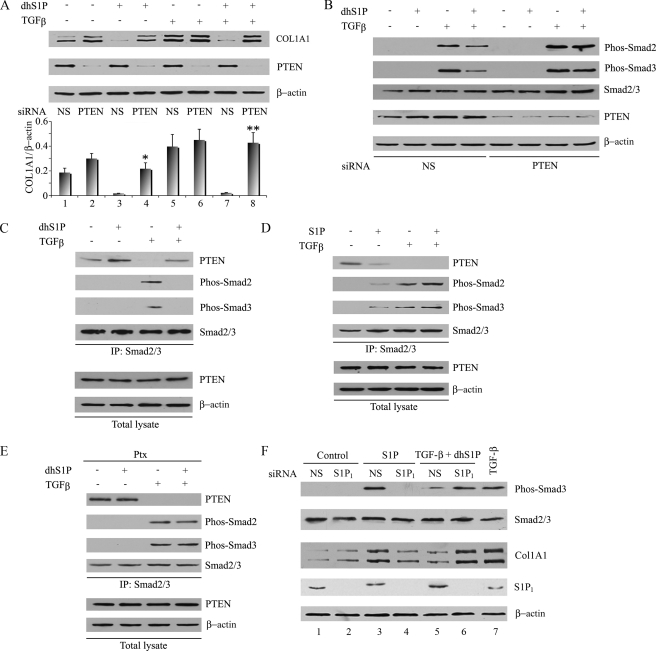
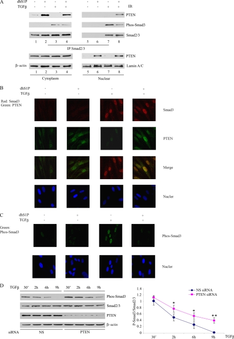
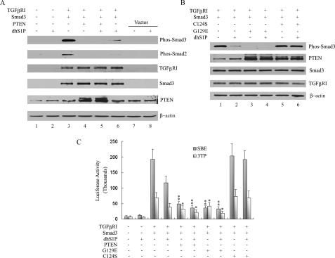
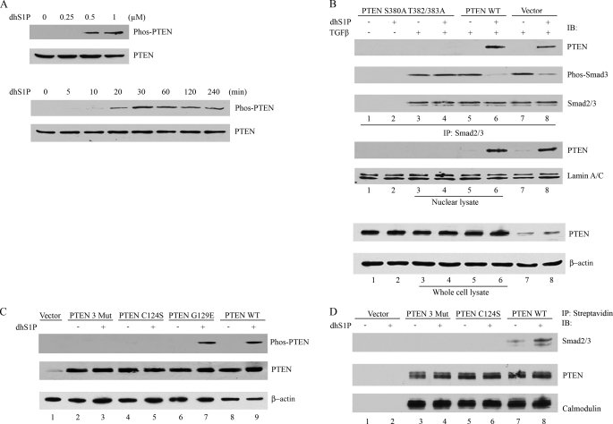
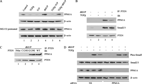
Transforming growth factor-beta (TGF-beta) is an important regulator of physiological connective tissue biosynthesis and plays a central role in pathological tissue fibrosis. Previous studies have established that a biologically active lipid mediator, sphingosine 1-phosphate (S1P), mimics some of the profibrotic functions of TGF-beta through cross-activation of Smad signaling. Here we report that another product of sphingosine kinase, dihydrosphingosine 1-phosphate (dhS1P), has an opposite role in the regulation of TGF-beta signaling. In contrast to S1P, dhS1P inhibits TGF-beta-induced Smad2/3 phosphorylation and up-regulation of collagen synthesis. The effects of dhS1P require a lipid phosphatase, PTEN, a key modulator of cell growth and survival. dhS1P stimulates phosphorylation of the C-terminal domain of PTEN and its subsequent translocation into the nucleus. We demonstrate a novel function of nuclear PTEN as a co-factor of the Smad2/3 phosphatase, PPM1A. Complex formation of PTEN with PPM1A does not require the lipid phosphatase activity but depends on phosphorylation of the serine/threonine residues located in the C-terminal domain of PTEN. Upon complex formation with PTEN, PPM1A is protected from degradation induced by the TGF-beta signaling. Consequently, overexpression of PTEN abrogates TGF-beta-induced Smad2/3 phosphorylation. This study establishes a novel role for nuclear PTEN in the stabilization of PPM1A. PTEN-mediated cross-talk between the sphingolipid and TGF-beta signaling pathways may play an important role in physiological and pathological TGF-beta signaling.


Figures

FIGURE 1.
FIGURE 1.
DhS1P and S1P have antagonistic effects on collagen production and Smad2/3 signaling. A, the collagen protein level was analyzed by Western blot in fibroblasts incubated with 0.5 μm dhS1P or 1 μm S1P and/or 2.5 ng/ml TGF-β for 24 h. The bar graph shows means ± S.E. of at least three independent experiments. *, dhS1P-treated versus control (p < 0.01); **, TGF-β + dhS1P versus TGF-β alone-treated cells (p < 0.01). B, fibroblasts were incubated with 2.5 ng/ml TGF-β for 30 min with the addition of the indicated concentrations of dhS1P (top) or incubated with S1P (bottom). Phosphorylated Smad2 was detected by Western blot. C, fibroblasts were treated with 2.5 ng/ml TGF-β and either 0.5 μm dhS1P or 1 μm S1P for 30 min. Phosphorylated Smad3 was detected by Western blot. D, HEK 293 cells were treated with 2.5 ng/ml TGF-β and 0.5 μm dhS1P for 30 min. Phosphorylated Smad2 and Smad3 were detected by Western blot. E, overexpression of SK1 prevents TGF-β-induced phosphorylation of Smad3. Fibroblasts were transduced with 50 multiplicity of infection of SK1 or control adenovirus (G0) for 24 h. Next, increasing concentration (0.5–2.5 ng/ml) of TGF-β was added for 30 min. Phosphorylated Smad3, total Smad2/3, and SK1 were detected by Western blot. β-Actin was used as loading control.
FIGURE 2.
FIGURE 2.
PTEN mediates inhibitory effects of dhS1P. A and B, depletion of endogenous PTEN prevents collagen down-regulation and Smad2/3 dephosphorylation by dhS1P. Fibroblasts were transfected with 30 nm PTEN siRNA or nonsilencing (NS) siRNA for 24 h and then serum-starved overnight. Cells were treated with 2.5 ng/ml TGF-β with the addition of 0.5 μm dhS1P for 30 min. Collagen 1A1 (A) and phosphorylated Smad2 and -3 (B) were detected by Western blot. The bar graph shows means ± S.E. of at least three independent experiments. *, dhS1P plus PTEN siRNA-treated versus dhS1P plus NS siRNA-treated cells (p < 0.01). **, TGF-β plus dhS1P and PTEN siRNA-treated versus TGF-β plus dhS1P and NS siRNA-treated cells (p < 0.001). C and D, dhS1P enhances whereas S1P diminishes binding of endogenous Smad2/3 and PTEN. Cells were treated with 2.5 ng/ml TGF-β with the addition of 0.5 μm dhS1P (C) or S1P (D) for 30 min. Smad2/3 was immunoprecipitated from cell extracts with anti-Smad2/3 antibody, followed by immunoblotting with PTEN, phospho-Smad2, and phospho-Smad3 antibodies. E, Ptx inhibits formation of Smad-PTEN complexes. Cells were pretreated with 100 ng/ml Pxt for 24 h and then treated with 0.5 μm dhS1P plus 2.5 ng/ml TGF-β for 30 min. Smad2/3 was immunoprecipitated from cell extracts, followed by immunoblotting with the indicated antibodies. F, depletion of endogenous S1P1 abrogates effects of S1P and dhS1P. Cells were transfected with 30 nm S1P1 siRNA or nonsilencing siRNA for 24 h and then serum-starved overnight. Cells were treated with 1 μm S1P or 2.5 ng/ml TGF-β plus 0.5 μm dhS1P for 30 min to measure phospho-Smad3 or 24 h to measure collagen. Phosphorylated Smad3 and COL1A1 protein were detected by immunoblotting. Cells treated with TGF-β only served as control (lane 7).
FIGURE 3.
FIGURE 3.
PTEN is required for Smad3 dephosphorylation in the nucleus. A, cytoplasmic and nuclear fractions were isolated from fibroblasts treated for 30 min with 2.5 ng/ml TGF-β and 0.5 μm dhS1P alone or in combination. Binding of PTEN to phosphorylated and total Smad2/3 were examined by immunoprecipitation (IP)/Western blot (IB). Distribution of PTEN in cytoplasmic and nuclear fractions was examined by immunoblotting. Lamin A/C and β-actin were used as control for nuclear and cytoplasmic fractions, respectively. B, immunofluorescence imaging of Smad3 (red) and PTEN (green) in cells treated individually or in combination with 0.5 μm dhS1P and 2.5 ng/ml TGF-β for 30 min. Note the co-localization of Smad3 and PTEN in cells treated by a combination of TGF-β and dhS1P. C, subcellular distribution of phosphorylated Smad3 (green) under experimental conditions described in B. Note the significantly reduced presence of nuclear phospho-Smad3 in cells treated with TGF-β and dhS1P. D, depletion of endogenous PTEN prolongs TGF-β-induced phosphorylation of Smad3. Cells were transfected with 30 nm PTEN siRNA or nonsilencing siRNA for 24 h and then starved overnight and incubated with 2.5 ng/ml TGF-β for the indicated times. The lower panel presents average kinetics of phospho-Smad3 from three independent experiments. *, p < 0.05; **, p < 0.01.
FIGURE 4.
FIGURE 4.
PTEN mediates dephosphorylation of Smad2/3 in a lipid phosphatase-independent manner. A, HEK 293 cells were co-transfected with the indicated combination of TGF-βRI 204D, Smad3, and PTENWT for 24 h and, where indicated, treated with 0. 5 μm dhS1P for 30 min. Expression levels of phospho-Smad3, phospho-Smad2, TGF-βRI, Smad3, and PTEN were assessed by immunoblotting. B, HEK 293 cells were cotransfected with the indicated combination of TGF-βRI 204D, Smad3, PTEN C124S, and PTEN G129E for 24 h and then treated with 0.5 μm dhS1P. Phospho-Smad3, PTEN, Smad3, and TGF-βRI were detected by immunoblotting. C, HEK 293 cells were transiently transfected with TGF-βRI 204D, Smad3, PTENWT, PTEN C124S, PTEN G129RE, and 3TP-Lux or SBE-Lux for 24 h and then incubated with 0.5 μm dhS1P for 30 min. Cells were harvested, and luciferase activity was measured. Activity is expressed as relative luminometer units (n ≥ 3). *, p < 0.05; **, p < 0.01.
FIGURE 5.
FIGURE 5.
C-terminal phosphorylation of PTEN mediates dhS1P-induced PTEN nuclear translocation and dephosphorylation of Smad3. A, dhS1P stimulates C-terminal phosphorylation of PTEN in a dose-dependent (top) and time-dependent (bottom) manner. C-terminal phosphorylation of PTEN was detected by immunoblotting. B, HEK 293 cells were transfected with the phosphorylation-deficient PTEN mutant, PTENWT, or vector only for 24 h and then incubated with 0.5 μm dhS1P and 2.5 ng/ml TGF-β for 30 min. The nuclear fraction was immunoprecipitated (IP) by Smad2/3 and then probed for PTEN, phospho-Smad3, and Smad2/3 by Western blot (IB). PTEN was also tested in nuclear lysate and whole cell lysate. Lamin A/C and β-actin served as controls for nuclear and cytoplasmic fractions, respectively. C, HEK 293 cells were transfected with the phosphorylation-deficient PTEN mutant (Mut3), PTEN C124S, PTEN G129E, and PTENWT and incubated with 0.5 μm dhS1P for 30 min. C-terminal phosphorylation of PTEN was detected by Western blotting. D, C-terminal phosphorylation (Mut3) and C124S PTEN mutants do not associate with Smad2/3. The PTEN proteins contained calmodulin and streptavidin tags from the pCTAP vector (Stratagene). Streptavidin-coupled agarose beads were used to pull down wild-type and mutated PTEN. Smad2/3 and PTEN were detected by immunoblotting. Immunoblotting with calmodulin served as control for equal loading.
FIGURE 6.
FIGURE 6.
Nuclear PTEN interacts with PPM1A and prevents its degradation by TGF-β. A, TGF-β reduces whereas dhS1P enhances PPM1A protein levels. Fibroblasts were incubated for 30 min with 2.5 ng/ml TGF-β and 0.5 μm dhS1P alone or in combination. PPM1A was detected by Western blot. The lower panel shows PPM1A levels under the same conditions in cells pretreated with 20 μm MG132 for 1 h. B, fibroblasts were treated for 30 min with 2.5 ng/ml TGF-β and 0.5 μm dhS1P alone or in combination. Binding of PTEN to PPM1A was examined by immunoprecipitation (IP)/Western blot (IB). C, HEK 293 cells were transfected with the phosphorylation-deficient PTEN mutant (Mut3), PTEN C124S, PTEN G129E, and PTENWT and incubated with 0.5 μm dhS1P for 30 min. Binding of PTEN to PPM1A was examined by immunoprecipitation/Western blot. D, depletion of endogenous PPM1A prevents inhibitory effects of PTEN on TGF-β-induced phosphorylation of Smad3. Cells were transfected with 30 nm PPM1A siRNA or nonsilencing siRNA for 24 h and then starved overnight and incubated for 30 min with 2.5 ng/ml TGF-β and 0.5 μm dhS1P alone or in combination. Smad3 and PPM1A were detected by Western blotting.

Similar articles

Cited by

References

    1. Massague, J. (1998) Annu. Rev. Biochem. 67 753-791 - PubMed
    1. Bierie, B., and Moses, H. L. (2006) Nat. Rev. Cancer 6 506-520 - PubMed
    1. Bhowmick, N. A., Chytil, A., Plieth, D., Gorska, A. E., Dumont, N., Shappell, S., Washington, M. K., Neilson, E. G., and Moses, H. L. (2004) Science 303 848-851 - PubMed
    1. Faler, B. J., Macsata, R. A., Plummer, D., Mishra, L., and Sidawy, A. N. (2006) Perspect. Vasc. Surg. Endovasc. Ther. 18 55-62 - PubMed
    1. Verrecchia, F., and Mauviel, A. (2007) World J. Gastroenterol. 13 3056-3062 - PMC - PubMed

Publication types

MeSH terms