Review
. 2008 May 15;22(10):1276-312.
doi: 10.1101/gad.1653708.

Role of platelet-derived growth factors in physiology and medicine

Affiliations
Review

Role of platelet-derived growth factors in physiology and medicine

Johanna Andrae et al. Genes Dev. .

Abstract

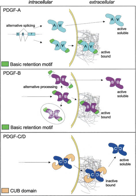
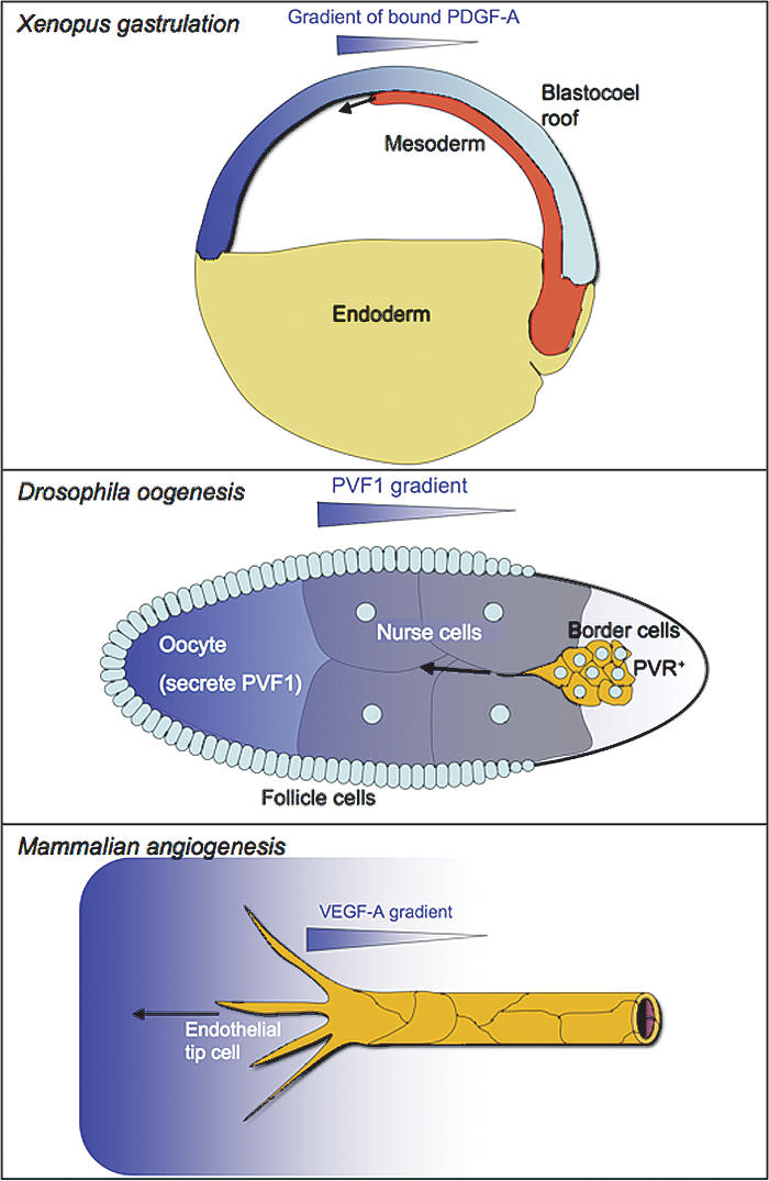
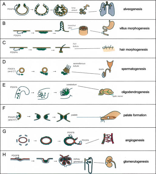
Platelet-derived growth factors (PDGFs) and their receptors (PDGFRs) have served as prototypes for growth factor and receptor tyrosine kinase function for more than 25 years. Studies of PDGFs and PDGFRs in animal development have revealed roles for PDGFR-alpha signaling in gastrulation and in the development of the cranial and cardiac neural crest, gonads, lung, intestine, skin, CNS, and skeleton. Similarly, roles for PDGFR-beta signaling have been established in blood vessel formation and early hematopoiesis. PDGF signaling is implicated in a range of diseases. Autocrine activation of PDGF signaling pathways is involved in certain gliomas, sarcomas, and leukemias. Paracrine PDGF signaling is commonly observed in epithelial cancers, where it triggers stromal recruitment and may be involved in epithelial-mesenchymal transition, thereby affecting tumor growth, angiogenesis, invasion, and metastasis. PDGFs drive pathological mesenchymal responses in vascular disorders such as atherosclerosis, restenosis, pulmonary hypertension, and retinal diseases, as well as in fibrotic diseases, including pulmonary fibrosis, liver cirrhosis, scleroderma, glomerulosclerosis, and cardiac fibrosis. We review basic aspects of the PDGF ligands and receptors, their developmental and pathological functions, principles of their pharmacological inhibition, and results using PDGF pathway-inhibitory or stimulatory drugs in preclinical and clinical contexts.


Figures

Figure 1.
Figure 1.
The PDGF/VEGF family in mammals and invertebrates. Mammalian PDGFs fall into two classes (I and II) distinguished by the presence of basic retention motifs (A and B) or CUB domains (C and D). Mammalian VEGFs also fall into two classes (III and IV). C. elegans (Ce) and Drosophila (D) PVFs are most similar to VEGF-C and VEGF-D based on domain organization but may functionally be most similar to VEGF-A, VEGF-B, and PlGF.
Figure 2.
Figure 2.
PDGF–PDGFR interactions. Each chain of the PDGF dimer interacts with one receptor subunit. The active receptor configuration is therefore determined by the ligand dimer configuration. The top panel shows the interactions that have been demonstrated in cell culture. Hatched arrows indicate weak interactions or conflicting results. The bottom panel shows interactions proven to be of importance in vivo during mammalian development. Note that PDGF-D has not yet been investigated in this regard.
Figure 3.
Figure 3.
Processing and extracellular retention of the PDGFs. (Top panel) Through alternative splicing, PDGF-A may be translated into a protein with or without a retention motif (green). Both isoforms may bind and activate PDGFR-α (active). Heterodimers between the long and short forms of PDGF-A are not illustrated but can likely form since both splice isoforms are produced by the same cells in most situations. (Middle panel) PDGF-B is produced as a single precursor containing the retention motif. This protein may be secreted, in which case it gets trapped at the external side of the cell membrane or in pericellular matrix such as the basement membrane, where it is active and participates in pericyte recruitment. In platelets, PDGF-B is processed intracellularly into a soluble and active isoform lacking the retention motif. There is experimental evidence for trafficking of a proportion of synthesized PDGF-B toward degradation without prior secretion. (Bottom panel) PDGF-C and PDGF-D are produced and secreted as inactive growth factors containing a CUB domain (yellow). The CUB domain may help in localizing these PDGFs in the extracellular space. Active PDGF-C and PDGF-D are produced through extracellular proteolysis.
Figure 4.
Figure 4.
PDGFR signal transduction and links to the cytoskeleton. (A) The intracellular domains of PDGFR-α and PDGFR-β and some of their direct interactors are illustrated. Arrows imply links to major signal transduction pathways and secondary effectors. Negative feedback signaling is indicated in red. (B) Schematic illustration of how PDGFR-β may link to the cytoskeleton and to other signaling components of focal adhesions through the adapter NHERF, the merlin and ezrin/radixin/moezin family of cytoskeletal linkers (MERM), and focal adhesion kinase (FAK).
Figure 5.
Figure 5.
Principles of pharmacological inhibition of PDGF signaling. PDGFs can be blocked by neutralizing antibodies, recombinant dimeric soluble PDGFR extracellular domains, or nucleic acids (aptamers), which all bind to the PDGFs with high affinity. PDGFRs can be blocked by antibodies or dominant-negative ligands. PDGFR function may be blocked by kinase inhibitors. The possibility of silencing the PDGF-A promoter by help of small-molecule pharmacologicals has emerged recently.
Figure 6.
Figure 6.
PDGFs, PVFs, and VEGFs direct cell migration in developmental processes. (Top panel) PDGF-A (long splice version) is expressed by the ectodermal cells of the blastocoel roof and gets deposited in a gradient that drives mesodermal cell movements along the blastocoel roof. (Middle panel) PVF1 is expressed by the oocyte and distributes in a graded fashion in the Drosophila egg chamber. This guides migration of the border cells toward the oocyte. (Bottom panel) Graded distribution of VEGF-A directs filopodial extensions and migration of the endothelial tip cells, thereby orienting the angiogenic sprout toward the highest VEGF-A concentration.
Figure 7.
Figure 7.
Developmental roles of PDGFs. (A) During the saccular phase of lung development, PDGF-A secreted by the epithelium (yellow) drives spreading and proliferation of alveolar SMC progenitors (green). These cells differentiate into alveolar SMC and drive formation and maintain alveolar walls by producing deposits of elastin. (B) In intestinal development, the epithelium produces PDGF-A to drive proliferation of mesenchymal cells with a critical role in intestinal villus formation. (C) In the developing skin, PDGF-A secreted from keratinocytes promote proliferation of mesenchymal cells with functions in hair follicle morphogenesis. (D) In testicular development, PDGF-A (and PDGF-C) secreted from the epithelial tubes drives expansion of mesenchymal cells that subsequently differentiate into testosterone-producing Leydig cells. The testosterone production is in turn necessary for further testicular development and spermatogenesis. (E) In the developing CNS (the example illustrates the cerebellum), PDGF-A drives proliferative expansion of OPs, which subsequently myelinate nerve fibers throughout the CNS. Lack of PDGF-A leads to a complete absence of myelin in peripheral parts of the CNS, such as the optic nerve. (F) PDGF-C (and PDGF-A) is critical for development of the palatal shelves. PDGF-C is produced in the epithelium and acts on mesenchymal cells (green) in the shelves. (G) PDGF-B produced by endothelial cells (orange) drives proliferation and spreading of vSMC and pericytes in conjunction with angiogenesis and arteriogenesis. (H) In developing kidney glomeruli, PDGF-B expressed by glomerular endothelium drives the proliferation of mesangial cells (green).
Figure 8.
Figure 8.
PDGFs in tumor biology. PDGFs are frequently produced by tumor cells and may affect tumor growth and dissemination in several different ways. PDGFs may directly (thick arrows) stimulate tumor cell growth and EMT. PDGFs may further be involved in the recruitment of tumor fibroblasts and pericytes. Tumor fibroblasts may in turn produce factors that directly act on the tumor cells to promote their proliferation and migration (HGF, CXCL12, FGF2, and FGF7). Tumor fibroblasts may also secrete angiogenic factors that help in sustaining tumor angiogenesis. Tumor fibroblasts may also promote metastasis by secreting CCL5, which induces metastatic behavior of the tumor cells. Tumor cell-derived PDGF-B and PDGF-D may, if produced in excess (such that the PDGF-B gradient established by the endothelial cells is overwhelmed), promote detachment of pericytes, which may facilitate metastasis. See the text for references to the different examples. The symbols are adapted from Hanahan and Weinberg (2000).
Figure 9.
Figure 9.
Genetic causes of PDGF/PDGFR deregulation. Translocation in the PDGF-B gene leads to an autocrine loop that sustains tumor cell proliferation in DP. Chromosomal translocations and deletions lead to the production of dimeric fusion proteins involving the intracellular PDGFR-β domain. Amplification of the PDGFR-A gene is frequently found in glioblastoma. GISTs may carry activating point mutations in the PDGFR-α kinase domain. See the text for details and references.
Figure 10.
Figure 10.
PDGFs and atherosclerosis. (A) Schematic illustration of a section through an arterial wall with an atherosclerotic plaque containing vSMC (green), inflammatory and immune cells (blue), and lipids (dark yellow). Invading inflammatory cells release a large number of growth factors and cytokines that maintain a chronic inflammatory reaction. PDGFs released by the inflammatory cells (and perhaps also from the endothelial cells) help in attracting vSMC from the media to the intima (light yellow). In a stable plaque, the intimal vSMC forms a subendothelial fibrous cap. Red arrows indicate routes of cell recruitment from the circulation and the arterial media. (B) LRP1 is a critical negative regulator of PDGFR-β in vSMC. This effect appears to be enhanced by ApoE. LRP1 inhibits PDGFR-β transcription (via TGF-β) and promotes PDGFR-β degradation (via c-Cbl). The interplay between LRP1 and PDGFR-β is complex. PDGF-B may bind to LRP1, and PDGFR-β phosphorylates LRP1.
Figure 11.
Figure 11.
PDGFs and fibrosis. (A) In lung fibrosis, PDGFs released from alveolar macrophages promote proliferation of alveolar (myo)fibroblasts and fibrogenesis. The latter express PDGFR-α, which is up-regulated by inflammatory cytokines, some of which are increased by air pollutants. (B) In liver fibrosis, hepatic stellate cells up-regulate PDGFR-β in response to TGF-β. PDGFs produced by resident and invading macrophages contribute to stellate cell proliferation and fibrogenesis. (C) In scleroderma, PDGFs released by macrophages promote proliferation of dermal (myo)fibroblasts. Fibroblast PDGFR-α and PDGF expression is up-regulated in response to Il-1α and TGF-β. Circulating autoantibodies that activate PDGF receptor have been described in scleroderma patients. (D) In a variety of renal diseases and disease models, PDGF-B released from invading macrophages and PDGF-D released from mesangial cells may drive mesangial cell proliferation and matrix deposition, leading to glomerulosclerosis. (E) In transgenic models, the expression of PDGFs in myocardial cells drives proliferative expansion of PDGFR-α-positive cardiac fibroblasts and collagen deposition, leading to severe cardiac fibrosis.

References

    1. Aase K., Abramsson A., Karlsson L., Betsholtz C., Eriksson U. Expression analysis of PDGF-C in adult and developing mouse tissues. Mech. Dev. 2002;110:187–191. - PubMed
    1. Abdollahi A., Li M., Ping G., Plathow C., Domhan S., Kiessling F., Lee L.B., McMahon G., Gröne H.J., Lipson K.E., et al. Inhibition of platelet-derived growth factor signaling attenuates pulmonary fibrosis. J. Exp. Med. 2005;201:925–935. - PMC - PubMed
    1. Abramsson A., Berlin O., Papayan H., Paulin D., Shani M., Betsholtz C. Analysis of mural cell recruitment to tumor vessels. Circulation. 2002;105:112–117. - PubMed
    1. Abramsson A., Lindblom P., Betsholtz C. Endothelial and nonendothelial sources of PDGF-B regulate pericyte recruitment and influence vascular pattern formation in tumors. J. Clin. Invest. 2003;112:1142–1151. - PMC - PubMed
    1. Abramsson A., Kurup S., Busse M., Yamada S., Lindblom P., Schallmeiner E., Stenzel D., Sauvaget D., Ledin J., Ringvall M., et al. Defective N-sulfation of heparan sulfate proteoglycans limits PDGF-BB binding and pericyte recruitment in vascular development. Genes & Dev. 2007;21:316–331. - PMC - PubMed

Publication types

MeSH terms

Substances

LinkOut - more resources