

doi: 10.1101/gad.469408.
Kindlin-2 controls bidirectional signaling of integrins
Affiliations
- PMID: 18483218
- PMCID: PMC2377186
- DOI: 10.1101/gad.469408
Item in Clipboard
Kindlin-2 controls bidirectional signaling of integrins
Genes Dev.
.
Abstract
Control of integrin activation is required for cell adhesion and ligand-induced signaling. Here we report that loss of the focal adhesion protein Kindlin-2 in mice results in peri-implantation lethality caused by severe detachment of the endoderm and epiblast from the basement membrane. We found that Kindlin-2-deficient cells were unable to activate their integrins and that Kindlin-2 is required for talin-induced integrin activation. Furthermore, we demonstrate that Kindlin-2 is required for integrin outside-in signaling to enable firm adhesion and spreading. Our findings provide evidence that Kindlin-2 is a novel and essential element of bidirectional integrin signaling.
Figures
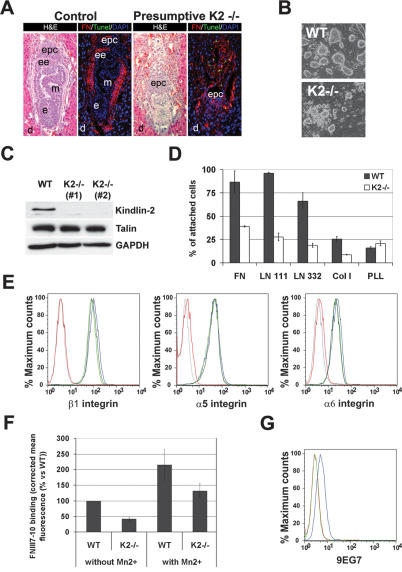
Peri-implantation lethality and impaired integrin activation in ESCs in the absence of Kindlin-2 expression. (A) Hematoxilin and eosin staining of an E6.5 control and presumptive Kindlin-2−/− implantation chamber. TUNEL staining (green) and FN staining (red) show apoptosis and disturbed matrix deposition in Kindlin-2−/− embryos. Nuclei are counterstained with DAPI (blue). (B) Bright-field pictures of wild-type and Kindlin-2−/− ESCs lines seeded on feeder cells. (C) Western Blot for Kindlin-2 and talin in wild-type and two independent Kindlin-2−/− ESCs lines. (D) Adhesion assay of wild-type and Kindlin-2−/− ESCs on different ECM substrates. (E) Integrin surface expression of wild-type (blue), Kindlin-2−/− (light and dark green), and β1-integrin−/− (red) ESC lines. A background control is shown in gray. (F) Binding of FNIII7-10 to wild-type and Kindlin-2−/− ESCs in the presence and absence of MnCl2. (G) 9EG7 binding on wild-type (blue) and Kindlin-2−/− (green) ESC lines. A background control is shown in red. (d) Decidua; (epc) ectoplacental cone; (ee) extraembryonic ectoderm; (e) ectoderm; (m) mesoderm.
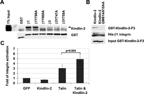
Kindlin-2 interacts with integrin tails and is required for integrin activation. (A) GST pull-down assay from ESC lysates with wild-type and mutant GST-tagged β1- and β3-integrin cytoplasmic tails. (B) Pull-down assays with the recombinant GST-tagged wild-type and mutant (QW614/615AA) PTB domain of Kindlin-2 and the recombinant His-tagged β1-integrin cytoplasmatic tail. (C) αIIbβ3-integrin activation in CHO cells transfected with EGFP, EGFP-Kindlin-2, EGFP-Talin head, and EGFP-Kindlin-2 together with the Talin head.
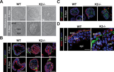
Kindlin-2 EBs show endoderm and epiblast detachment and diminished integrin activation. (A) Micrographs of methylene blue-stained sections of 4- and 6-d-old wild-type and Kindlin-2−/− EBs. (B) Cryosections of wild-type and Kindlin-2−/− EBs were stained with an antibody specific for LN-α1 chain (green) and fluorescently labeled phalloidin to visualize F-actin (red). Nuclei are counterstained with DAPI (blue). (C) TUNEL staining (green), LN-α1 chain (red), and nuclei (blue) of 6-d-old wild-type and Kindlin-2−/− EBs. (D) Six-day-old wild-type and Kindlin-2−/− EBs stained with LN-α1 chain (green) and 9EG7 (red) antibodies. Nuclei are counterstained with DAPI (blue). (epi) Epiblast; (end) endoderm.
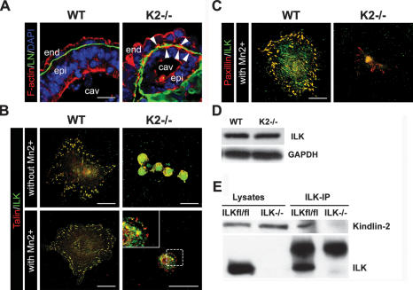
Impaired cell spreading of Kindlin-2−/− endoderm cells. (A) Cryosections of 6-d-old wild-type and Kindlin-2−/− EBs stained with LN-α1 chain antibody (green) and fluorescently labeled phalloidin to visualize F-actin (red). Nuclei are counterstained with DAPI (blue). (B) Immunofluorescence staining for talin (red) and ILK (green) of wild-type and Kindlin-2−/− endoderm cells seeded of FN in the presence or absence of MnCl2. (C) Immunofluorescence staining for paxillin (red) and ILK (green) of wild-type and Kindlin-2−/− endoderm cells seeded of FN in the presence of MnCl2. (D) Western blot analysis of ILK in wild-type and Kindlin-2−/− endoderm cells. (E) Coimmunoprecipitation of ILK and Kindlin-2 from ILKfl/fl and ILK−/− fibroblasts.
References
-
- Aumailley M., Pesch M., Tunggal L., Gaill F., Fässler R. Altered synthesis of laminin 1 and absence of basement membrane component deposition in β1 integrin-deficient embryoid bodies. J. Cell Sci. 2000;113:259–268. - PubMed
-
- Calderwood D.A., Zent R., Grant R., Rees D.J., Hynes R.O., Ginsberg M.H. The Talin head domain binds to integrin β subunit cytoplasmic tails and regulates integrin activation. J. Biol. Chem. 1999;274:28071–28074. - PubMed
-
- Calderwood D.A., Yan B., de Pereda J.M., Alvarez B.G., Fujioka Y., Liddington R.C., Ginsberg M.H. The phosphotyrosine binding-like domain of talin activates integrins. J. Biol. Chem. 2002;277:21749–21758. - PubMed
-
- Chen J., Salas A., Springer T.A. Bistable regulation of integrin adhesiveness by a bipolar metal ion cluster. Nat. Struct. Biol. 2003;10:995–1001. - PubMed
Publication types
MeSH terms
Substances
Grants and funding
LinkOut - more resources
Full Text Sources
Other Literature Sources
Molecular Biology Databases