

doi: 10.1101/gad.463608.
Control of final seed and organ size by the DA1 gene family in Arabidopsis thaliana
Affiliations
- PMID: 18483219
- PMCID: PMC2377187
- DOI: 10.1101/gad.463608
Item in Clipboard
Control of final seed and organ size by the DA1 gene family in Arabidopsis thaliana
Genes Dev.
.
Abstract
Although the size of an organism is a defining feature, little is known about the mechanisms that set the final size of organs and whole organisms. Here we describe Arabidopsis DA1, encoding a predicted ubiquitin receptor, which sets final seed and organ size by restricting the period of cell proliferation. The mutant protein encoded by the da1-1 allele has a negative activity toward DA1 and a DA1-related (DAR) protein, and overexpression of a da1-1 cDNA dramatically increases seed and organ size of wild-type plants, identifying this small gene family as important regulators of seed and organ size in plants.
Figures
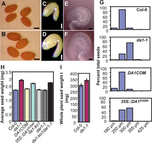
Seed size in da1-1. (A,B) Dry seeds of Col-0 (A) and da1-1 (B). (C,D) Mature embryos of Col-0 (C) and da1-1(D). (E,F) Ovules of Col-0 (E) and da1-1 (F). (G) Preweighed batches of wild-type Col-0, da1-1, complementation line (DA1COM#2), and 35S∷DA1R358K #5 seeds from individual plants were passed through a series of wire sieves of decreasing mesh size (in microns) as described in the Supplemental Material. Seeds retained by each sieve were weighed, and the mass of each fraction was expressed as a percentage of the total mass of the seed sample analyzed. (H) Average seed weights of Col-0, da1-1, DA1COM#2, 35S∷DA1R358K #5, da1-ko1, dar1-1, and da1-ko1dar1-1 double mutants are shown as milligrams per 100 seeds. Standard deviations are shown (n = 5). (I) Average seed weight per plant. Standard deviation values were given (n = 5). Plants were grown under identical conditions. Bars: A,B, 200 μm; C,D, 100 μm; E,F, 50 μm.
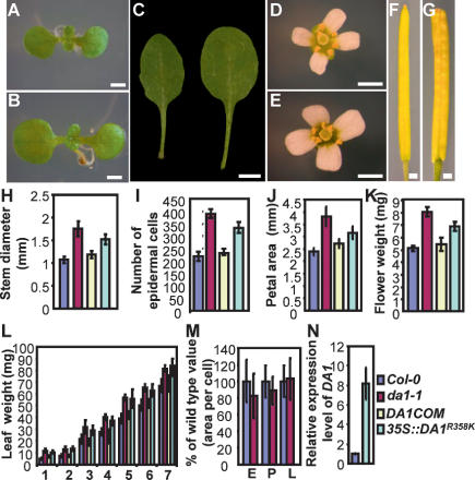
Organ size in da1-1. (A,B) (A) Nine-day-old seedlings of Col-0 (A) and da1-1 (B). da1-1 has larger cotyledons than wild type. (C) The fifth leaves of Col-0 (left) and da1-1 (right). da1-1 has larger and rounder leaves compared with wild-type Col-0. (D,E) Flowers of Col-0 (D) and da1-1 (E). (F,G) Siliques of Col-0 (F) and da1-1 (G). (H–J) Stem diameter (H), epidermal cell number in stem cross-sections (I), and petal area (J) of Col-0, da1-1, DA1COM#2, and 35S∷DA1R358K#5. (K,L) Mass of five fresh flowers (stage 14) (K) and leaves (first through seventh) of 35-d-old plants (L). (M) Cell area of embryos (E), petals (P), and leaves (L) in Col-0 and da1-1. Values are given as mean ± SD relative to the respective wild-type value, set at 100%. (N) Relative expression levels of DA1 in Col-0 and 35S∷DA1R358K#5 seedlings were measured by quantitative real-time RT–PCR. Bars: A,B, 1 mm; C, 0.5 cm; D–G, 1 mm.
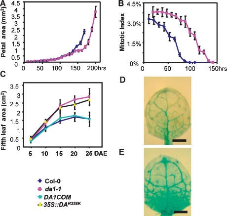
Kinematic analysis of petal and leaf growth. (A) Growth of Col-0 and da1-1 mutant petals. The largest petals of each series are from opened flowers (stage 14). (B) Mitotic index in wild-type and da1-1 mutant petals. The time axis in B corresponds to that in A. (C) Fifth leaf area of Col-0, da1-1, DA1COM#2, and 35S∷DA1R358K#5 measured over 25 d. (DAE) Days after emergence. (D,E) pCyclinB1;1∷GUS activity in the first leaves (9 d after germination) of wild-type (D) and da1-1 (E) seedlings grown on MS medium containing 1% glucose. Bar, 1 mm.
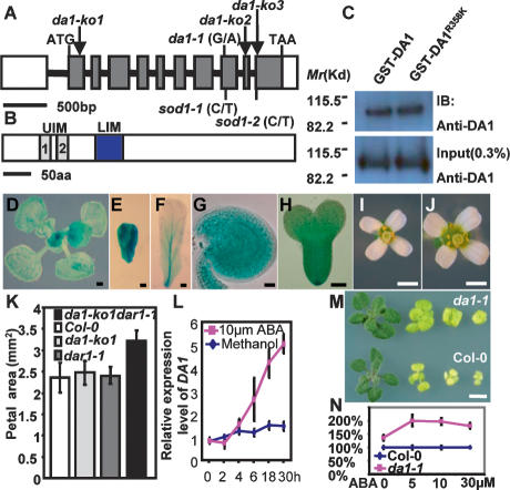
Identification, expression, function, and genetic interactions of the DA1 gene. (A) DA1 gene structure showing the mutated sites of da1-1, sod1-1, and sod1-2 alleles. The start codon (ATG) and the stop codon (TAA) are indicated. Closed boxes indicate the coding sequence, and lines between boxes indicate introns. T-DNA insertion sites (da1-ko1, da1-ko2, and da1-ko3) in DA1 gene are shown. (B) The predicted DA1 protein contains two UIM and one LIM domains. (C) In vitro ubiquitin-binding assay. GST-DA1 and GST-DA1R358K fusion proteins expressed in E. coli were incubated with ubiquitin–agarose beads. Bound GST-DA1 and GST-DA1R358K fusion proteins (top panel) and 0.3% input proteins (bottom panel) were detected by immunoblotting (IB) with an antibody against DA1 (anti-DA1). (D–H) DA1 expression activity was monitored by pDA1∷GUS transgene expression. GUS staining during early leaf development (D), developing (E) and mature (F) petals, a developing ovule (G), and early-stage embryo (H). (I,J) Flowers of Col-0 (I) and da1-ko1dar1-1 double mutant (J). (K) Petal area of Col-0, da1-ko1, dar1-1, and da1-ko1dar1-1 double mutants. The da1-ko1dar1-1 double mutant displays a da1-1 phenotype including large flowers and petals. (L) Quantitative RT–PCR analysis revealed that expression of DA1 is slowly induced by ABA. Seven-day-old wild-type seedlings were treated with 10 μM ABA for 2, 4, 6, 18, and 30 h. (M) Seedlings of Col-0 (bottom) and da1-1 (top) were established on MS medium for 4 d and transferred to MS + 1% glucose medium without ABA and with 5 μm ABA, 10 μM ABA, and 30 μM ABA (from left to right) for 2 wk, respectively. The da1-1 mutant seedlings exhibit ABA-insensitive organ growth, compared with Col-0. (N) The shoot fresh weight of Col-0 and da1-1 in M. Values are given as mean ± SE relative to the respective wild-type value, set at 100%. Bars: D,M, 0.5 mm; E,F, 0.1 mm; G,H, 50 μm; I,J, 1 mm; (O, 2 mm.
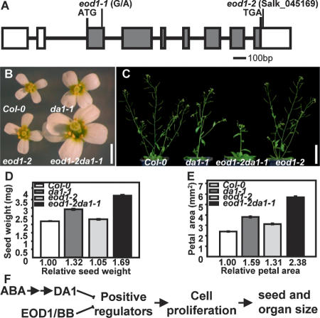
Mutations in EOD1/BB synergistically enhance the phenotypes of da1-1. (A) EOD1/BB gene structure, showing the mutated sites of the two eod1 alleles. The start codon (ATG) and the stop codon (TGA) are indicated. Closed boxes indicate the coding sequence, and lines between boxes indicate introns. The mutated site in eod1-1 and T-DNA insertion site in eod1-2 also are shown. (B) Flowers of Col-0, da1-1, eod1-2, and eod1-2da1-1 double mutants. (C) Soil-grown plants of Col-0, da1-1, eod1-2da1-1 double mutant, and eod1-2 are shown. (D) Average seed weights of Col-0, da1-1, eod1-2, and eod1-2da1-1 double mutants are shown as milligrams per 100 seeds. Standard deviations are shown (n = 5). Plants were grown under identical conditions. (E) Petal areas of Col-0, da1-1, eod1-2, and eod1-2da1-1 double mutant. Standard deviation values are shown (n > 50). (F) A model of DA1 and EOD1/BB controlling seed and organ size. Bars: B, 2 mm; C, 50 mm.
References
-
- Conlon I., Raff M. Size control in animal development. Cell. 1999;96:235–244. - PubMed
-
- Dawid I.B., Breen J.J., Toyama R. LIM domains: Multiple roles as adapters and functional modifiers in protein interactions. Trends Genet. 1998;14:156–162. - PubMed
-
- Day S.J., Lawrence P.A. Measuring dimensions: The regulation of size and shape. Development. 2000;127:2977–2987. - PubMed
-
- Dinneny J.R., Yadegari R., Fischer R.L., Yanofsky M.F., Weigel D. The role of JAGGED in shaping lateral organs. Development. 2004;131:1101–1110. - PubMed
Publication types
MeSH terms
Substances
LinkOut - more resources
Full Text Sources
Other Literature Sources
Molecular Biology Databases