. 2008 May 15;22(10):1345-55.
doi: 10.1101/gad.470008.

Coordinated regulation of transcriptional repression by the RBP2 H3K4 demethylase and Polycomb-Repressive Complex 2

Affiliations

Coordinated regulation of transcriptional repression by the RBP2 H3K4 demethylase and Polycomb-Repressive Complex 2

Diego Pasini et al. Genes Dev. .

Abstract

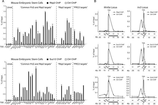
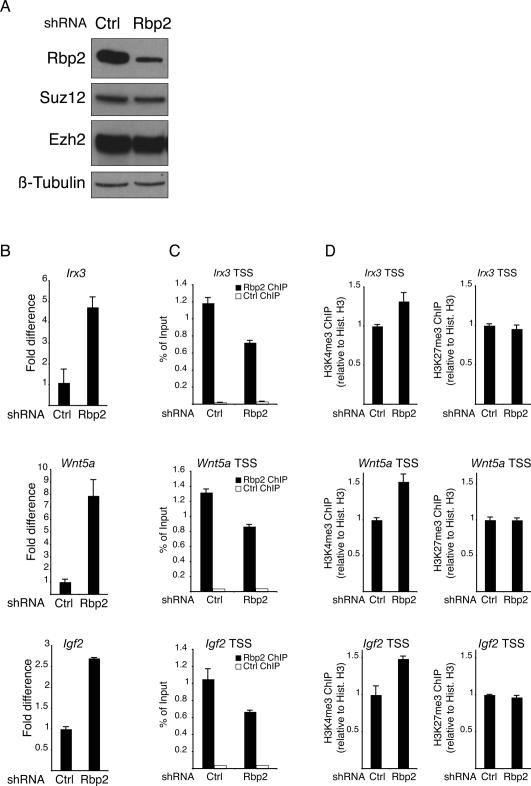
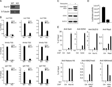
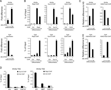
Polycomb group (PcG) proteins regulate important cellular processes such as embryogenesis, cell proliferation, and stem cell self-renewal through the transcriptional repression of genes determining cell fate decisions. The Polycomb-Repressive Complex 2 (PRC2) is highly conserved during evolution, and its intrinsic histone H3 Lys 27 (K27) trimethylation (me3) activity is essential for PcG-mediated transcriptional repression. Here, we show a functional interplay between the PRC2 complex and the H3K4me3 demethylase Rbp2 (Jarid1a) in mouse embryonic stem (ES) cells. By genome-wide location analysis we found that Rbp2 is associated with a large number of PcG target genes in mouse ES cells. We show that the PRC2 complex recruits Rbp2 to its target genes, and that this interaction is required for PRC2-mediated repressive activity during ES cell differentiation. Taken together, these results demonstrate an elegant mechanism for repression of developmental genes by the coordinated regulation of epigenetic marks involved in repression and activation of transcription.


Figures

Figure 1.
Figure 1.
Identification of Rbp2 target genes in mouse ES cells by genome-wide location analysis. (A) ChIP assays in control and RBP2 shRNA-treated 293T cells on the BRD8 promoter. ChIP with antibody against the hemagglutinin (HA) tag served as negative control. ChIP enrichments are presented as percentage (%) of bound/input signal. (B) Clustering of Rbp2-bound promoters in mouse ES cells into functional groups. (C) Comparison of PcG target genes and their regulatory pathways published elsewhere (Bracken et al. 2006) with those regulated by Rbp2. (D) Statistically significant overlap between promoters bound by Rbp2 and promoters bound by PcGs published elsewhere (Boyer et al. 2006) in ES cells. The P-value was calculated by hypergeometric probability test using Genespring software (Agilent).
Figure 2.
Figure 2.
Rbp2 and PRC2 occupy overlapping target genes. (A) ChIP analysis in ES cells using antibodies against Rbp2 and Suz12. The first group includes unrelated control genes, the second group includes genes found in both Rbp2 and Suz12 genome-wide location analysis, the third group provides genes found bound by Rbp2 only, and the fourth group of genes are bound by PRC2 only. Antibody against the HA tag served as negative control. ChIP enrichments are presented as percentage (%) of bound/input signal. (B) ChIP analysis in mouse ES cells of Rbp2, Suz12, and Ezh2 along the Wnt5a and Irx3 gene loci. ChIPs with antibody against the HA tag served as negative control. ChIP enrichments are presented as percentage (%) of bound/input signal. The position of the qPCR amplicons (arrows) relative to the genes (black boxes). TSS (dashed line) are also presented in the figure.
Figure 3.
Figure 3.
RBP2 interacts with the PRC2 complex. (A) IP assays using 293T cell lysates expressing HA-tagged wild-type (WT) or enzymatically inactive RBP2 mutant showing specific coimmunoprecipitation of RBP2 with EZH2 and EED. (B) IPs of mouse ES cell lysates using specific antibodies to Ezh2 and Suz12 showing specific coimmunoprecipitation of Rbp2. IP with anti-HA-tag antibody served as negative control. (C, top panels) Size exclusion chromatography (Superose-6, GE Healthcare) of mouse ES cell protein lysate showing coelution of Rbp2, Ezh2, Suz12, and Eed in high-molecular-weight fractions. (Bottom panels) The indicated fractions were immunoprecipitated with a Suz12-specific antibody. IP with anti-HA-tag antibody served as negative control (Ctrl). (D) Coomassie-stained gel and Western blot of protein-immunoprecipitated material from Sf9 cells expressing recombinant RBP2, EZH2, SUZ12, and Eed with anti-SUZ12 and anti-HA-tag (as negative control) antibodies showing the binding of RBP2 to the PRC2 complex.
Figure 4.
Figure 4.
Rbp2 is required for the repression of PRC2 target genes. (A) Western blot analysis of protein lysates prepared from mouse ES cells transduced with either control or Rbp2 shRNA lentiviruses. (BD) Expression levels of the Irx3, Wnt5a, and Igf2 (B) and ChIP assays of the Irx3, Wnt5a, and Igf2 promoters using antibodies specific for Rbp2, histone H3, H3K4me3, and H3K27me3 (C,D) in control (Ctrl) or Rbp2 shRNA-treated mouse ES cells. ChIPs with antibody against HA tag served as negative control. ChIP enrichments are presented as percentage (%) of bound/input signal. ChIPs for H3K4me3 and H3K27me3 are further normalized to histone H3 density. Gene expression is normalized to Gapdh levels.
Figure 5.
Figure 5.
The PRC2 complex is required for Rbp2 recruitment to target genes. (A) Western blots from Suz12 wild-type (WT) and knockout (KO) mouse ES cells using the indicated antibodies. β-Tubulin served as a loading control. (B) ChIP assays for Suz12, Ezh2, and Rbp2 on the Irx3, Wnt5a, and Igf2 promoters in wild-type (WT) and knockout (KO) ES cells. ChIPs with antibody against HA tag served as negative control. ChIP enrichments are presented as percentage (%) of bound/input signal. (C) Western blot analysis using antibodies specific for EED, SUZ12, and EZH2 of lysates prepared from a Gal4-EED-inducible 293T cell line grown with or without tetracycline. (D) Luciferase activity on cell lysates prepared from GAL4-EED 293T cells grown with or without tetracycline. (E) ChIP assays of lysates prepared from GAL4-EED 293T cells grown with or without tetracycline using the indicated antibodies. ChIP with antibody against HA tag served as negative control. ChIP enrichments are presented as percentage (%) of bound/input signal.
Figure 6.
Figure 6.
Regulation of Rbp2 and PRC2 activities during ES cell differentiation. (A) Real-time quantitative expression analysis of Wnt5a and Fgf4 in ES cells before and 48 h after ATRA-induced (1 μM) differentiation. (B) ChIP assay in pluripotent and differentiated ES cells presented in A for Rbp2, Suz12, and Ezh2. ChIPs with antibody against HA tag served as negative control. ChIP enrichments are presented as percentage (%) of bound/input signal. (C) ChIP analysis as described in A using antibodies for histone H3, H3K4me3, and H3K27me3. ChIP enrichments for H3K4me3 and H3K27me3 are normalized to histone H3 density. (D) ChIP assays for Suz12 and Rbp2 in mouse ES, MEF, and C2C12 cells. ChIPs with antibody against the HA tag served as negative control. ChIP enrichments are presented as percentage (%) of bound/input signal.
Figure 7.
Figure 7.
Models for Rbp2–PRC2-repressive mechanisms during ES cell differentiation.

References

    1. Agger K., Cloos P.A., Christensen J., Pasini D., Rose S., Rappsilber J., Issaeva I., Canaani E., Salcini A.E., Helin K. UTX and JMJD3 are histone H3K27 demethylases involved in HOX gene regulation and development. Nature. 2007;449:731–734. - PubMed
    1. Agger K., Christensen J., Cloos P.A., Helin K. The emerging functions of histone demethylases. Curr. Opin. Genet. Dev. 2008 doi: 10.1016/j.gde.2007.12.003. - DOI - PubMed
    1. Benevolenskaya E.V., Murray H.L., Branton P., Young R.A., Kaelin W.G. Binding of pRB to the PHD protein RBP2 promotes cellular differentiation. Mol. Cell. 2005;18:623–635. - PubMed
    1. Bernstein B.E., Mikkelsen T.S., Xie X., Kamal M., Huebert D.J., Cuff J., Fry B., Meissner A., Wernig M., Plath K., et al. A bivalent chromatin structure marks key developmental genes in embryonic stem cells. Cell. 2006;125:315–326. - PubMed
    1. Boyer L.A., Plath K., Zeitlinger J., Brambrink T., Medeiros L.A., Lee T.I., Levine S.S., Wernig M., Tajonar A., Ray M.K., et al. Polycomb complexes repress developmental regulators in murine embryonic stem cells. Nature. 2006;441:349–353. - PubMed

Publication types

MeSH terms