. 2008 Aug 1;112(3):822-9.
doi: 10.1182/blood-2008-03-142182. Epub 2008 May 15.

Truncation in CCND1 mRNA alters miR-16-1 regulation in mantle cell lymphoma

Affiliations

Truncation in CCND1 mRNA alters miR-16-1 regulation in mantle cell lymphoma

Robert W Chen et al. Blood. .

Abstract

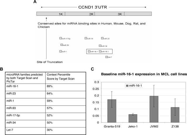
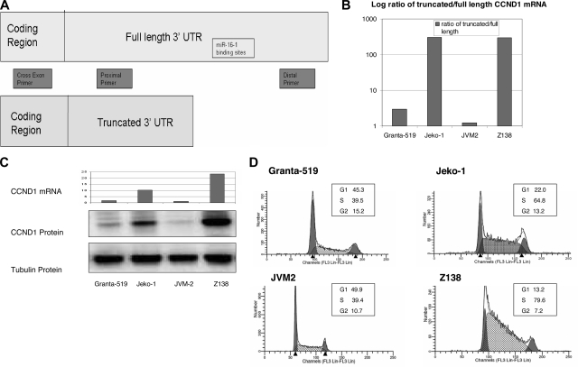
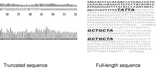
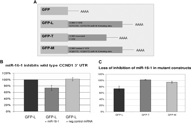
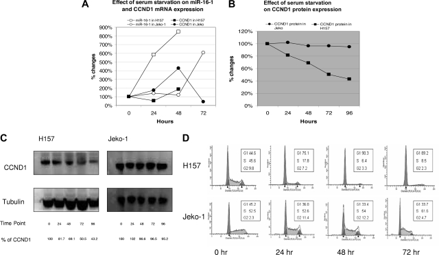
Cyclin D1 (CCND1) is a well-known regulator of cell-cycle progression. It is overexpressed in several types of cancer including breast, lung, squamous, neuroblastoma, and lymphomas. The most well-known mechanism of overexpression is the t(11;14)(q13;q32) translocation found in mantle cell lymphoma (MCL). It has previously been shown that truncated CCND1 mRNA in MCL correlates with poor prognosis. We hypothesized that truncations of the CCND1 mRNA alter its ability to be down-regulated by microRNAs in MCL. MicroRNAs are a new class of abundant small RNAs that play important regulatory roles at the posttranscriptional level by binding to the 3' untranslated region (UTR) of mRNAs blocking either their translation or initiating their degradation. In this study, we have identified the truncation in CCND1 mRNA in MCL cell lines. We also found that truncated CCND1 mRNA leads to increased CCND1 protein expression and increased S-phase cell fraction. Furthermore, we demonstrated that this truncation alters miR-16-1 binding sites, and through the use of reporter constructs, we were able to show that miR-16-1 regulates CCND1 mRNA expression. This study introduces the role of miR-16-1 in the regulation of CCND1 in MCL.


Figures

Figure 1
Figure 1
CCND1 is a target of miR-16-1 as predicted by Target Scan. (A) Shown are the seed binding regions located in the distal region of the CCND1 3′ UTR. The 2 binding sites for miR-16-1 are boxed. The truncation (shown by ↑) deletes CCND1 all miRNA binding sites. (Adapted from Target Scan.) (B) Table showing the context percentile score of each microRNA family predicted to target CCND1 by Target Scan. The higher the context score, the higher the probability that the microRNA will be able to inhibit CCND1 translation. (C) qRT-PCR to show the endogenous miR-16-1 expression in MCL cell lines. Error bars represent SD.
Figure 2
Figure 2
Truncated CCND1 leads to increased CCND1 mRNA, increased CCND1 protein expression, and cell-cycle progression. (A) Primer design for qRT-PCR to assess relative values of truncated versus full-length CCND1 mRNA. The cross exon primer set will amplify only mRNA in the coding region, thus avoiding genomic contamination. The proximal primer set will amplify both the full-length and truncated mRNA. The distal primer set will amplify only the full-length mRNA. (B) qRT-PCR showing the log ratio of truncated to full-length CCND1 mRNA in MCL cell lines. mRNA is expressed as relative quantitation equalized to beta-actin. Jeko-1 and Z138 had much higher quantitation values for truncated message and lower values for full-length message compared with Granta-519 and JVM2. Similarly, the ratio of truncated to full-length CCND1 mRNA is much higher in Jeko-1 and Z138. (C) Relative expression of total amount of CCND1 mRNA and corresponding CCND1 protein expression in 4 MCL cell lines. Cross exon primers were designed to amplify total CCND1 mRNA to avoid genomic contamination. Jeko-1 and Z138 also have increased total amount of CCND1 mRNA compared with Granta-519 and JVM-2. Similarly, Jeko-1 and Z-138 have increased CCND1 protein expression compared with Granta-519 and JVM-2. (D) Cell cycle in MCL cell lines. Jeko-1 and Z138 have increased percentage of cells in S-phase compared with Granta-519 and JVM-2.
Figure 3
Figure 3
Site of CCND1 mRNA truncation. NCode was used to clone the truncated CCND1 mRNA from MCL cell lines as previously described (“GFP-CCND1-3′ UTR reporter constructs”). Truncation occurs 345 bp downstream of the stop codon and eliminates both miR-16-1 binding sites. Note that the poly A tails immediately follow the sequence TATTA in sequencing data. The letters in gray reflect the truncated sequences, and GCTGCTA denotes miR-16-1 binding sites.
Figure 4
Figure 4
miR-16-1 regulates CCND1 through its 3′ UTR. (A) Three GFP reporter constructs were made as previously described (“Methods”). GFP-L contains the distal CCND1 3′ UTR including both miR-16-1 binding sites. GFP-T contains the truncated CCND1 3′ UTR. GFP-M is a double mutant where both miR-16-1 binding sites were mutated. Notice the mutation changes the T from the third base pair to an A and the A in the seventh base pair to a T. (B) H157 cells were transfected with GFP-L and cotransfected with a mimic of miR-16-1 or negative control random miRNA. Shown is the GFP expression measured by flow cytometry. RFP-expressing plasmids were cotransfected to control for transfection efficiency. Error bars represent SD.(C) H157 cells were transfected with either GFP-L, GFP-T, or GFP-M. They were also cotransfected with either a mimic of miR-16-1 or negative control random miRNA. GFP expression of cells transfected by a mimic of miR-16-1 is first normalized to the RFP expression and then normalized to cells transfected with negative control random miRNA. This shows that miR-16-1 mimic cannot regulate a truncated or mutated CCND1 3′ UTR.
Figure 5
Figure 5
Endogenous miR-16-1 inhibits CCND1 translation in H157. (A) Serum starvation increases miR-16-1 by approximately 800% in H157 and approximately 600% in Jeko-1. Serum starvation also increases CCND1 mRNA approximately 200% in H157 but decreases CCND1 mRNA in Jeko-1. (B) Serum starvation decreases CCND1 protein expression in H157 by approximately 30% at 48 hours and has no effect on CCND1 protein expression in Jeko-1 even at 96 hours. (C) Western blot analysis showing down-regulation of CCND1 protein expression in H157, while no changes occurred in Jeko-1 with serum starvation. (D) Cell-cycle analysis showing cell-cycle progression in H157 and Jeko-1 with serum starvation. The accumulation of cells in G1-phase in H157 at 48 hours indicates that serum starvation causes cell-cycle arrest. Notice that cell-cycle progression does not change dramatically in Jeko-1 even with serum starvation.

References

    1. Campo E, Raffeld M, Jaffe ES. Mantle-cell lymphoma. Semin Hematol. 1999;36:115–127. - PubMed
    1. Jemal A, Siegel R, Ward E, Murray T, Xu J, Thun MJ. Cancer statistics, 2007. CA Cancer J Clin. 2007;57:43–66. - PubMed
    1. Witzig TE. Current treatment approaches for mantle-cell lymphoma. J Clin Oncol. 2005;23:6409–6414. - PubMed
    1. Bertoni F, Rinaldi A, Zucca E, Cavalli F. Update on the molecular biology of mantle cell lymphoma. Hematol Oncol. 2006;24:22–27. - PubMed
    1. Quelle DE, Ashmun RA, Shurtleff SA, et al. Overexpression of mouse D-type cyclins accelerates G1 phase in rodent fibroblasts. Genes Dev. 1993;7:1559–1571. - PubMed

Publication types