. 2008 Jun 13;320(5882):1507-10.
doi: 10.1126/science.1159051. Epub 2008 May 15.

Activation of the cellular DNA damage response in the absence of DNA lesions

Affiliations

Activation of the cellular DNA damage response in the absence of DNA lesions

Evi Soutoglou et al. Science. .

Abstract

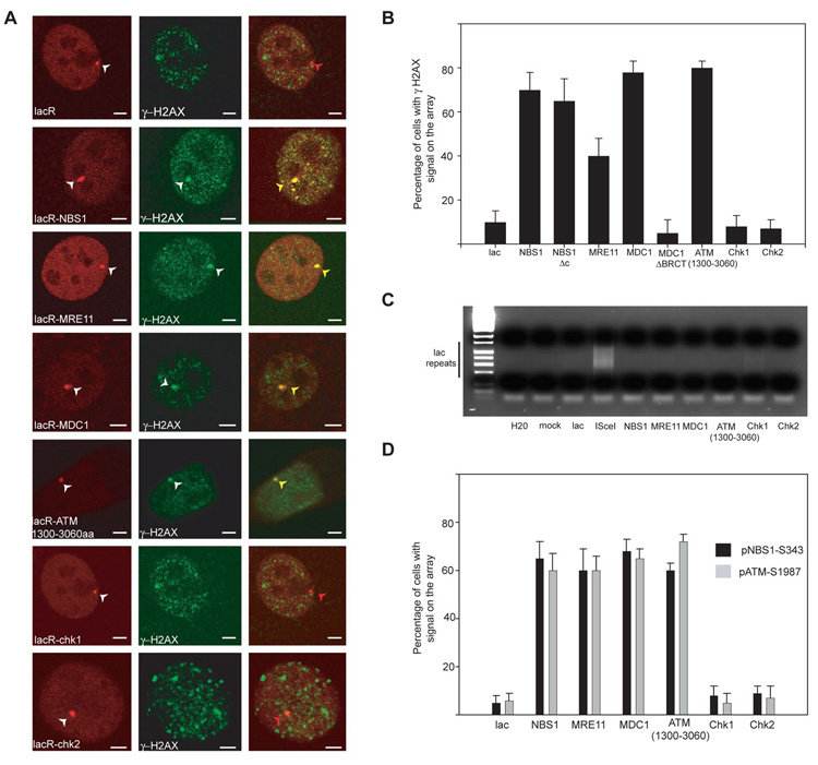
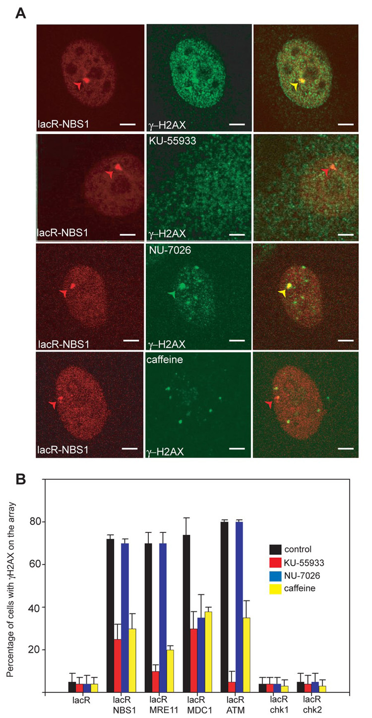
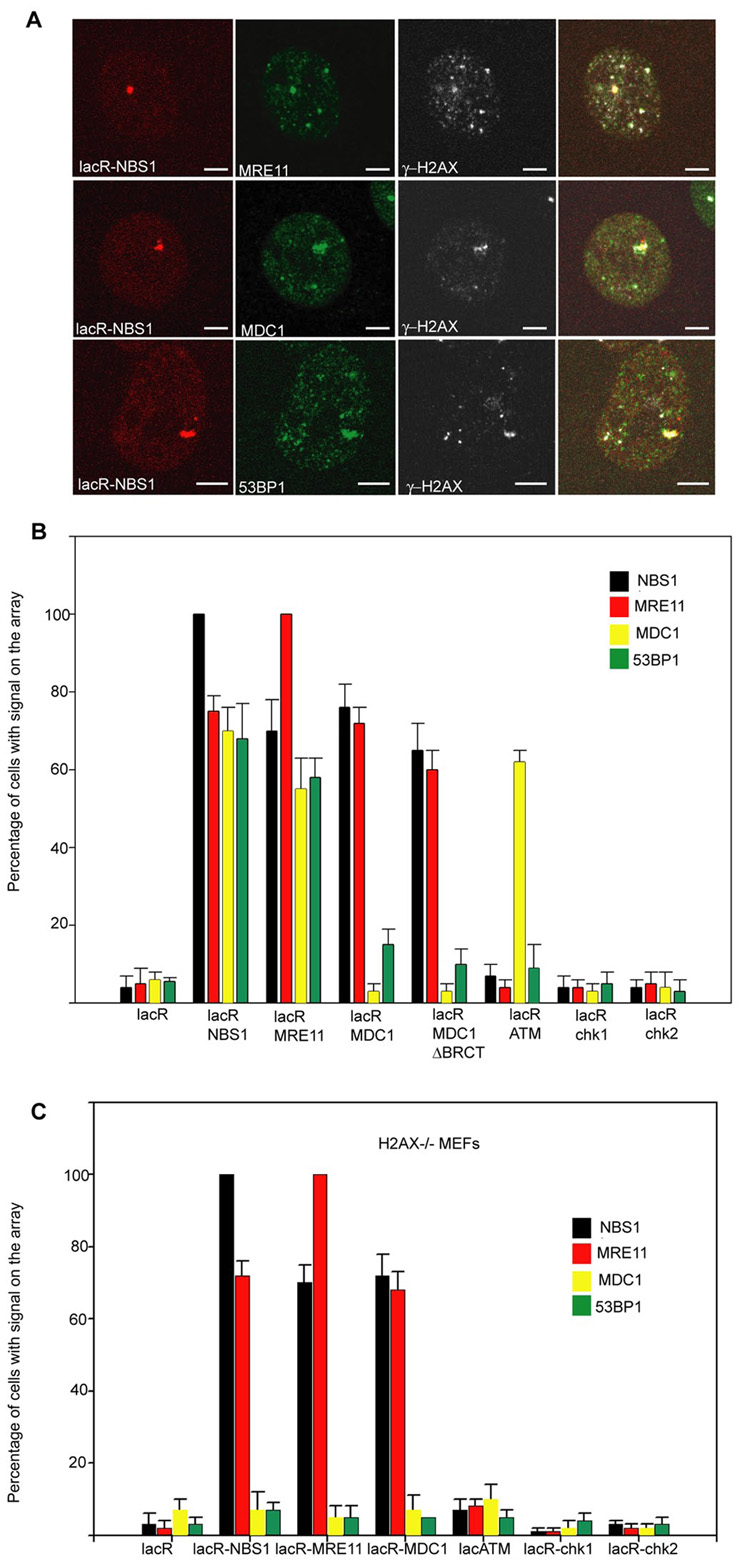
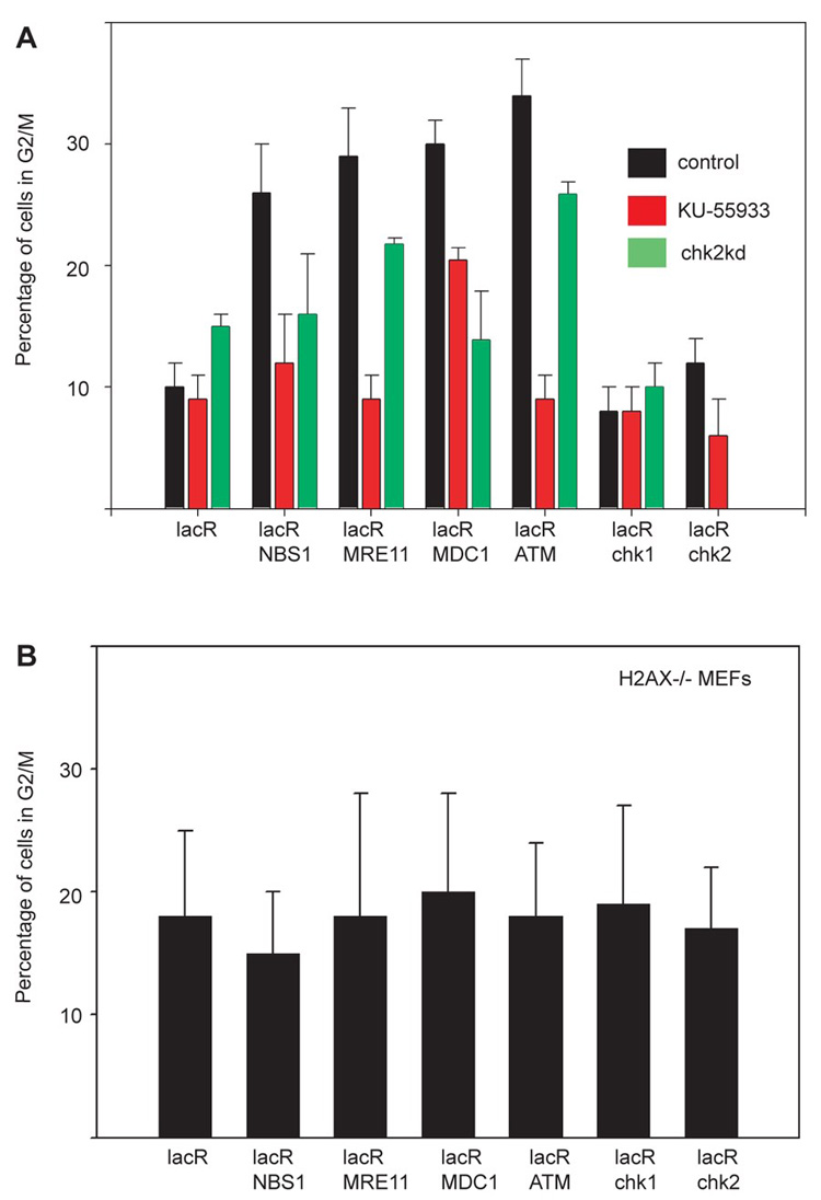
The cellular DNA damage response (DDR) is initiated by the rapid recruitment of repair factors to the site of DNA damage to form a multiprotein repair complex. How the repair complex senses damaged DNA and then activates the DDR is not well understood. We show that prolonged binding of DNA repair factors to chromatin can elicit the DDR in an ATM (ataxia telangiectasia mutated)- and DNAPK (DNA-dependent protein kinase)-dependent manner in the absence of DNA damage. Targeting of single repair factors to chromatin revealed a hierarchy of protein interactions within the repair complex and suggests amplification of the damage signal. We conclude that activation of the DDR does not require DNA damage and stable association of repair factors with chromatin is likely a critical step in triggering, amplifying, and maintaining the DDR signal.


Figures

Fig. 1
Fig. 1
Immobilization of single repair factors on chromatin leads to DDR activation. (A) Immunofluorescence microscopy on NIH 2/4 cells transiently transfected for 16h with the indicated repair factor fused to Cherry-LacR (red). DDR activation is indicated by phosphorylation of H2AX (green). Scale Bar = 2µm. (B) Quantitative analysis of H2AX phosphorylation on the lacO array after transfection of the indicated repair factors for 16h. Values represent averages ± S.D (n = 40) from 4 independent experiments. (C) Absence of DNA lesions upon tethering. Ligation-mediated PCR on NIH2/4 cells transfected for 16h with the indicated repair factor fused to Cherry-LacR. The PCR product is a ladder due of the repetitive nature of the lac operator. Transfection of ISceI alone is used as a control. (D) Quantitative analysis of NBS1 (S343) or ATM (S1987) phosphorylation on the array after transfection of the indicated repair factor for 24 h. Values represent averages ± S.D (n = 60) from 2 independent experiments.
Fig. 2
Fig. 2
Activation of DDR depends on ATM and DNAPK. (A) Detection of γH2AX by indirect immunofluorescence in NIH 2/4 cells transiently transfected for 24h with lacR-NBS1- CherryRed (red) and treated with the indicated kinase inhibitor for 24h. γH2AX (green). Scale Bar = 2µm. (B) Quantitative analysis of H2AX phosphorylation on the lacO array after immobilization of the indicated repair factors in the presence of signaling inhibitors. Values represent averages ± S.D (n = 50) from 4 independent experiments.
Fig. 3
Fig. 3
Cross-recruitment of repair factors. (A) Immunofluorescence microscopy on NIH 2/4 cells transiently transfected for 24h with lacR-NBS1 (red) with the indicated antibodies (green). Scale Bar = 2µm. (B) Quantitation of repair factor accumulation on the array in NIH 2/4 cells after immobilization of the indicated repair factors. Values represent averages ± S.D (n = 40) from 3 independent experiments. The antibody used to detect MDC1 recognizes only the mouse isoform and not human MDC1 present in the LacR fusion protein. (C) Quantitation of repair factor accumulation on the array on H2AX−/− MEFs containing the lacO array after immobilization of the indicated repair factors. Values represent averages ± S.D (n = 50) from 3 independent experiments.
Fig. 4
Fig. 4
Targeting of single repair factors to chromatin induces G2 delay. (A) Quantitation of cells with phosphoH3 in Ser10 on pericentromeric chromatin on NIH 2/4 cells after immobilization of the indicated repair factors in wt cells, cells treated with ATM inhibitor or cells transfected with siRNA against chk2 . The siRNA pool eliminated also the expression of the fusion protein (absence of green bar in LacR-Chk2). Values represent averages ± S.D (n = 150) from at least two independent experiments. (B) Quantitation of cells with phosphoH3 in Ser10 on pericentromeric chromatin on H2AX−/− MEFs after immobilization of the indicated repair factors. The baseline level of phosphoS10H3 is slightly elevated in MEFs compared to NIH 2/4 cells. Values represent averages ± S.D (n = 150) from at least two independent experiments

References

    1. Lukas J, Bartek J. Cell. 2004;118:666. - PubMed
    1. Shiloh Y. Curr Opin Genet Dev. 2001;11:71. - PubMed
    1. Jackson SP. Carcinogenesis. 2002;23:687. - PubMed
    1. Bekker-Jensen S, et al. J Cell Biol. 2006;173:195. - PMC - PubMed
    1. Bartek J, Lukas J. Cancer Cell. 2003;3:421. - PubMed

Publication types

MeSH terms

Substances