Review
. 2008 Jun;6(6):455-65.
doi: 10.1038/nrmicro1887.

Coordinating assembly of a bacterial macromolecular machine

Affiliations
Review

Coordinating assembly of a bacterial macromolecular machine

Fabienne F V Chevance et al. Nat Rev Microbiol. 2008 Jun.

Abstract

The assembly of large and complex organelles, such as the bacterial flagellum, poses the formidable problem of coupling temporal gene expression to specific stages of the organelle-assembly process. The discovery that levels of the bacterial flagellar regulatory protein FlgM are controlled by its secretion from the cell in response to the completion of an intermediate flagellar structure (the hook-basal body) was only the first of several discoveries of unique mechanisms that coordinate flagellar gene expression with assembly. In this Review, we discuss this mechanism, together with others that also coordinate gene regulation and flagellar assembly in Gram-negative bacteria.


Figures

Figure 1
Figure 1. Electron micrograph images illustrating the different types of flagellar arrangement in bacteria
A single flagellum can be present at one end of the cell (monotrichous); for example, in Vibrio cholerae, Pseudomonas aeruginosa, Idiomarina loihiensis (a) and Caulobacter crescentus (b). Many bacteria have numerous flagella and, if these are co-located on the surface of the cell to form a tuft, the bacterium is lophotrichous; for example, Vibrio fischeri (c) and Spirillum spp. Peritrichous flagella are distributed all over the cell; for example Escherichia coli and Salmonella enterica serovar Typhimurium (d). For spirochaetes, such as species of Borrelia (e), Treponema and Leptospira, a specialized set of flagella are located in the periplasmic space, the rotation of which causes the entire bacterium to move forward in a corkscrew-like motion. Images kindly provided by S.-I. Aizawa, Prefectural University of Hiroshima, Japan.
Figure 2
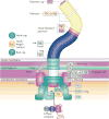
Figure 2. Flagellar components of Salmonella enterica serovar Typhimurium
Shows the structure of the bacterial flagellum as it resides within the cell wall and membranes (BOX 1). Soluble cytoplasmic components include the FliH–FliI–FliJ ATPase complex, which is thought to deliver a number of the secreted substrates and help determine the order of substrate secretion. The filament protein consists of either FliC or FljB, which are alternately transcribed. The rod cap (FlgJ) and the hook cap (FlgD) are transiently associated with the flagellum during rod and hook polymerization, respectively. FliK is secreted during rod–hook polymerization as a molecular ruler that couples rod–hook length to the flagellar secretion specificity switch at FlhB.
Figure 3
Figure 3. Coupling of flagellar gene regulation to flagellum assembly
The flhDC operon, or flagellar master operon, is under the control of numerous global regulatory signals that lead to the expression or inhibition of flagellar gene expression. Induction of the class I flhDC operon (class I on) produces FlhD and FlhC, which form a heteromultimeric complex, FlhD4C2, that acts to direct σ-dependent transcription from class II flagellar promoters and auto-repress flhDC transcription (class I off; class II on). Class II promoters direct the transcription of genes that are necessary for the structure and assembly of the hook–basal body (HBB) substructure. Upon HBB completion, late secretion substrates are exported from the cell and their cognate chaperones are released to regulate gene expression. FliT is an FlhD4C2 factor and prevents both FlhD4C2 auto-repression and the activation of class II promoters. The σ transcription factor directs the transcription of class III promoters, which include the filament structural genes and the genes of the chemosensory pathway (class I on; class II off; and class III on). Activation of class I transcription would re-initiate the flagellar regulon for a new round of flagellar gene expression. As drawn, the FliK and FlhA proteins are meant to reside within the C ring. The stoichiometries of Fluke, FlhA, FlhB, FliO, FliP, FliQ and FliR within the C ring are not known.

References

    1. Bardy SL, Ng SY, Jarrell KF. Recent advances in the structure and assembly of the archaeal flagellum. J Mol Microbiol Biotechnol. 2004;7:41–51. - PubMed
    1. Merz AJ, So M, Sheetz MP. Pilus retraction powers bacterial twitching motility. Nature. 2000;407:98–102. - PubMed
    1. Mignot T, Shaevitz JW, Hartzell PL, Zusman DR. Evidence that focal adhesion complexes power bacterial gliding motility. Science. 2007;315:853–856. - PMC - PubMed
    1. Seto S, Uenoyama A, Miyata M. Identification of a 521-kilodalton protein (Gli521) involved in force generation or force transmission for Mycoplasma mobile gliding. J Bacteriol. 2005;187:3502–3510. - PMC - PubMed
    1. Hiratsuka Y, Miyata M, Tada T, Uyeda TQ. A microrotary motor powered by bacteria. Proc Natl Acad Sci USA. 2006;103:13618–13623. - PMC - PubMed

Publication types

MeSH terms