Review
. 2008 Jun;8(6):421-34.
doi: 10.1038/nri2322.

The regulation of IgA class switching

Affiliations
Review

The regulation of IgA class switching

Andrea Cerutti. Nat Rev Immunol. 2008 Jun.

Abstract

IgA class switching is the process whereby B cells acquire the expression of IgA, the most abundant antibody isotype in mucosal secretions. IgA class switching occurs via both T-cell-dependent and T-cell-independent pathways, and the antibody targets both pathogenic and commensal microorganisms. This Review describes recent advances indicating that innate immune recognition of microbial signatures at the epithelial-cell barrier is central to the selective induction of mucosal IgA class switching. In addition, the mechanisms of IgA class switching at follicular and extrafollicular sites within the mucosal environment are summarized. A better understanding of these mechanisms may help in the development of more effective mucosal vaccines.


Conflict of interest statement

Competing interests statement

The author declares no competing financial interests.

Figures

Figure 1
Figure 1. Recombinatorial and transcriptional events underlying IgA class switching
The immunoglobulin heavy chain (IgH) locus of mature B cells contains a rearranged variable (V) diversity (D) joining (J) exon encoding the antigen-binding domain of an immunoglobulin. Following rearrangement of the light chain, B cells produce intact IgM and IgD through a transcriptional process driven by a promoter (P) upstream of the VDJ exon. Production of downstream IgG, IgA or IgE with identical antigen specificity occurs through class-switch recombination (CSR). Appropriate stimuli induce germline transcription of the constant heavy chain α (Cα) gene from the promoter (Pα) of the intronic α (Iα) exon through the switch α (Sα) region between Iα and Cα exons. In addition to yielding a sterile Iα–Cα mRNA, germline transcription renders the Cα gene substrate for activation-induced cytidine deaminase (AID), an essential component of the CSR machinery. By generating and repairing DNA breaks at Sμ and Sα, the CSR machinery rearranges the IgH locus, thereby yielding a deletional recombination product known as the switch circle. This episomal DNA transcribes a chimeric Iα–Cμ mRNA under the influence of signals that activate Pα. Post-switch transcription of the IgH locus generates mRNAs for both secreted IgA and membrane IgA. Cα 1–3, exons that encode the Cα chain of IgA; S, 3′ portion of Cα3 encoding the tailpiece of secreted IgA; M, exon encoding the transmembrane and cytoplasmic portions of membrane-bound IgA; αs, polyadenylation site for secreted IgA mRNA; αm, polyadenylation site for membrane-bound IgA mRNA.
Figure 2
Figure 2. Signalling events leading to T-cell-dependent IgA class switching
CD4+ T cells release the active transforming growth factor-β1 (TGFβ1) after processing of a latency-associated peptide (LAP). TGFβ1 forms a heteromeric TGFβ receptor (TGFβR) complex on B cells comprising TGFβRII and TGFβRI subunits. TGFβR undergoes degradation on binding by I-SMAD (inhibitory SMAD (mothers against decapentaplegic homologue)) proteins, such as SMAD7, which recruits ubiquitin ligases of the SMURF (SMAD ubiquitylation regulatory factor) family to TGFβRI. Alternatively, the TGFβR remains on the B-cell surface to activate SMAD proteins. In the presence of TGFβ1, TGFβRII kinases phosphorylate TGFβRI, leading to the activation of TGFβRI kinases. These kinases induce the phosphorylation of receptor-regulated SMAD (R-SMAD) proteins, including SMAD2 and SMAD3, thereby releasing them from the plasma membrane-anchoring protein SARA (SMAD anchor for receptor activation). After forming homo-oligomeric complexes, as well as hetero- oligomeric complexes with SMAD4 — a co-mediator SMAD (Co-SMAD) protein — R-SMAD proteins translocate to the nucleus, where they bind to SMAD-binding elements (SBEs) on target gene promoters, including constant heavy chain α (Cα) gene promoters. These SMAD complexes further associate with constitutive and TGFβR-induced co-factors, including runt-related transcription factor 3 (RUNX3), which binds to RUNX-binding elements (RBEs), cyclic AMP response element binding protein (CREB), which binds to a cyclic AMP response element (CRE), and Ets-like factor 1 (ELF1), which binds to an Ets-binding site. In addition to TGFβ, CD4+ T cells express CD40 ligand (CD40L), which elicits oligomerization of CD40 on B cells, recruitment of tumour-necrosis-factor-receptor-associated factors (TRAFs) to CD40, activation of the IκB kinase (IKK) complex, phosphorylation of IκB (inhibitor of nuclear factor-κB (NF-κB)), and IκB degradation. IκB-free NF-κB translocates to the nucleus to induce the activation-induced cytidine deaminase (AICDA) gene promoter. Although NF-κB binds to an NF-κB-binding (κB) site on the Cα promoter, it has a marginal role in the transcription of the Cα gene.
Figure 3
Figure 3. Signalling events leading to T-cell-independent IgA class switching
Dendritic cells (DCs) activate transforming growth factor-β1 (TGFβ1) by inducing the processing of a latency-associated peptide (LAP). TGFβ1 activates the constant heavy chain α (Cα) gene promoter (as shown in Figure 2). DCs also present bacterial products to B cells, thereby activating Toll-like receptors (TLRs). By recruiting myeloid differentiation primary-response protein 88 (MyD88), interleukin-1-receptor-associated kinase 1 (IRAK1) and IRAK4, TLRs induce activation of the IκB (inhibitor of nuclear factor-κB (NF-κB)) kinase (IKK) complex, phosphorylation and degradation of IκB. IκB-free NF-κB translocates to the nucleus to induce the promoter of the activation-induced cytidine deaminase (AICDA) gene. DCs further activate B cells by engaging transmembrane activator and calcium-modulating cyclophilin-ligand interactor (TACI) through B-cell-activating factor (BAFF) and a proliferation-inducing ligand (APRIL). TACI activates NF-κB after recruiting tumour-necrosis-factor-receptor-associated factors (TRAFs) to its cytoplasmic domain, thereby triggering AICDA gene expression. It is unknown whether TLRs and TACI also activate the Cα promoter. Co-SMAD, co-mediator SMAD; CRE, cyclic AMP response element; CREB, CRE-binding protein; ELF1, Ets-like factor 1; κB, NF-κB-binding site; RUNX3, runt-related transcription factor 3; RBE, RUNX-binding element; SARA, SMAD anchor for receptor activation; SBE, SMAD-binding element; SMAD, mothers against decapentaplegic homologue; TGFβR, TGFβ1 receptor.
Figure 4
Figure 4. Map of IgA class switching in the gut
Dendritic cells (DCs) in the subepithelial dome (SED) of the Peyer’s patches capture antigen by interacting with microfold (M) cells or by extending transepithelial projections into the lumen. During this process, DCs are induced to express tumour-necrosis factor (TNF) and inducible nitric oxide synthase (iNOS) (and are therefore referred to as tiDCs), which present antigen to perifollicular CD4+ T cells, thereby inducing them to differentiate into effector T cells releasing IgA-inducing cytokines. T cells also interact with antigen-specific IgM+IgD+ naive B cells. Together with follicular dendritic cells (FDCs), this interaction fosters a germinal centre (GC) reaction that includes somatic hypermutation (SHM) and IgA class-switch recombination (CSR). The resulting IgA+ effector B cells home to the gut lamina propria, where they differentiate into plasma cells that secrete high-affinity IgA. Human IgA1+ effector B cells can also undergo sequential IgA2 CSR on receiving T-cell-independent signals from bacteria-activated epithelial cells, DCs and tiDCs. Similar signals trigger direct IgA CSR in various B-cell subsets, including unmutated IgM+IgD+ B-1 cells from the peritoneum and mutated IgM+IgD effector B cells from Peyer’s patches. These local CSR events generate plasma cells secreting low- or high-affinity IgA. FAE, follicle-associated epithelium; FM, follicular mantle; HEV, high endothelial venule; sIgA, secreted IgA.
Figure 5
Figure 5. Cellular interactions causing IgA class switching in the gut
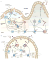
a | While capturing antigen, intestinal dendritic cells (DCs) are exposed to microbial Toll-like receptor (TLR) ligands and epithelial-cell-derived cytokines, including thymic stromal lymphopoietin (TSLP). These signals promote the generation of tiDCs, which are DCs that express tumour-necrosis factor and inducible nitric oxide synthase; these cells present antigen to CD4+ T cells in the perifollicular area of Peyer’s patches. In addition, tiDCs transfer antigen to follicular IgM+IgD+ naive B cells and induce them to upregulate the expression of TGFβ1 receptor (TGFβR) through nitric oxide (NO). During cognate interactions with CD4+ T cells, B cells undergo IgA class-switch recombination (CSR) in response to CD40 ligand (CD40L) and transforming growth factor-β1 (TGFβ1) from activated T cells. IgA expression requires interleukin-5 (IL-5), IL-6 and IL-10 from activated T cells, as well as B-cell-activating factor (BAFF) and a proliferation-inducing ligand (APRIL) from tiDCs. After being imprinted by retinoic acid (RA) from DCs, IgA+ effector B cells migrate to the lamina propria, where they differentiate into IgA-secreting plasma cells. This differentiation is enhanced by APRIL secreted by epithelial cells, DCs and tiDCs. b | After sensing microorganisms via TLRs, epithelial cells from intestinal villi release APRIL, thereby triggering direct IgA CSR in lamina-propria IgM+ B cells and sequential IgA2 CSR in lamina-propria IgA1+ B cells in a T-cell-independent manner. This pathway may also involve APRIL, BAFF and TGFβ1 from DCs and tiDCs exposed to microbial TLR ligands, NO and TSLP. Epithelial cells, DCs and tiDCs promote plasma-cell differentiation via APRIL, BAFF, IL-6 and IL-10. BCMA, B-cell maturation antigen; FAE, follicle-associated epithelium; M cell, microfold cell; pIgR, polymeric immunoglobulin receptor; SED, subepithelial dome; sIgA, secreted IgA; TACI, transmembrane activator and calcium-modulating cyclophilin-ligand interactor.

References

    1. Macpherson AJ, McKoy KD, Johansen FE, Brandtzaeg P. The immune geography of IgA induction and function. Mucosal Immunol. 2008;1:11–22. An overview of the function and topography of IgA responses and the mechanisms of their induction. - PubMed
    1. Fagarasan S, Honjo T. Intestinal IgA synthesis: regulation of front-line body defences. Nature Rev Immunol. 2003;3:63–72. - PubMed
    1. Macpherson AJ, Harris NL. Interactions between commensal intestinal bacteria and the immune system. Nature Rev Immunol. 2004;4:478–485. - PubMed
    1. Schlissel MS. Regulating antigen-receptor gene assembly. Nature Rev Immunol. 2003;3:890–899. - PubMed
    1. Odegard VH, Schatz DG. Targeting of somatic hypermutation. Nature Rev Immunol. 2006;6:573–583. - PubMed

Publication types

MeSH terms

Substances