. 2008 May 16;4(5):e1000066.
doi: 10.1371/journal.ppat.1000066.

The uptake of apoptotic cells drives Coxiella burnetii replication and macrophage polarization: a model for Q fever endocarditis

Affiliations

The uptake of apoptotic cells drives Coxiella burnetii replication and macrophage polarization: a model for Q fever endocarditis

Marie Benoit et al. PLoS Pathog. .

Expression of concern in

Abstract

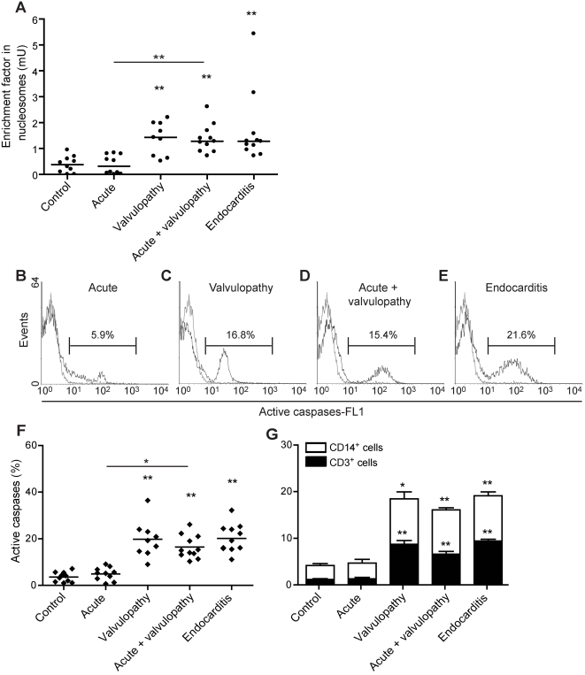
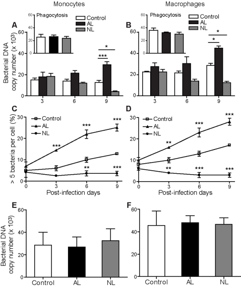
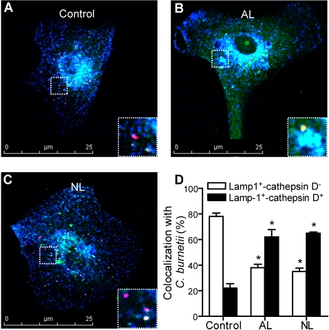
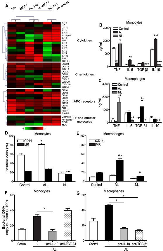
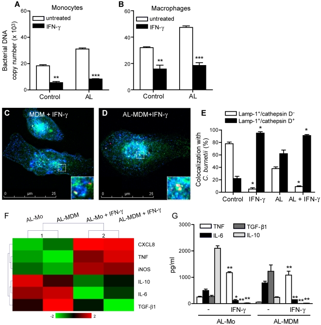
Patients with valvulopathy have the highest risk to develop infective endocarditis (IE), although the relationship between valvulopathy and IE is not clearly understood. Q fever endocarditis, an IE due to Coxiella burnetii, is accompanied by immune impairment. Patients with valvulopathy exhibited increased levels of circulating apoptotic leukocytes, as determined by the measurement of active caspases and nucleosome determination. The binding of apoptotic cells to monocytes and macrophages, the hosts of C. burnetii, may be responsible for the immune impairment observed in Q fever endocarditis. Apoptotic lymphocytes (AL) increased C. burnetii replication in monocytes and monocyte-derived macrophages in a cell-contact dependent manner, as determined by quantitative PCR and immunofluorescence. AL binding induced a M2 program in monocytes and macrophages stimulated with C. burnetii as determined by a cDNA chip containing 440 arrayed sequences and functional tests, but this program was in part different in monocytes and macrophages. While monocytes that had bound AL released high levels of IL-10 and IL-6, low levels of TNF and increased CD14 expression, macrophages that had bound AL released high levels of TGF-beta1 and expressed mannose receptor. The neutralization of IL-10 and TGF-beta1 prevented the replication of C. burnetii due to the binding of AL, suggesting that they were critically involved in bacterial replication. In contrast, the binding of necrotic cells to monocytes and macrophages led to C. burnetii killing and typical M1 polarization. Finally, interferon-gamma corrected the immune deactivation induced by apoptotic cells: it prevented the replication of C. burnetii and re-directed monocytes and macrophages toward a M1 program, which was deleterious for C. burnetii. We suggest that leukocyte apoptosis associated with valvulopathy may be critical for the pathogenesis of Q fever endocarditis by deactivating immune cells and creating a favorable environment for bacterial persistence.


Conflict of interest statement

The authors have declared that no competing interests exist.

Figures

Figure 1
Figure 1. Circulating apoptosis markers in patients with valvulopathy.
A, The circulating levels of nucleosomes was determined by immunoassays in control subjects, patients with acute Q fever, valvulopathy, acute Q fever with valvulopathy and Q fever endocarditis. Results are expressed as individual values with medians. B–E, Active caspases were detected through FLICA fluorescence (FL1 channel) and flow cytometry analysis of leukocytes from patients with acute Q fever (B), valvulopathy (C), acute Q fever with valvulopathy (D) and Q fever endocarditis (E) compared to control subjects (gray line). F, The results are expressed as percentage of active caspases for each individual. Bars represent the median of the values. G, Active caspases were quantified in CD3+ and CD14+ leukocytes from control subjects and patients. The results represent the mean±SEM. * p<0.01 and ** p<0.001.
Figure 2
Figure 2. Binding of AL and NL to Mo and MDM.
A and B, Mo (A) and MDM (B) were incubated with AL and NL for 30 min. Mo and MDM were fixed, gold-coated and analyzed by SEM or labeled with BODIPY phallacidin and observed in fluorescence microscopy. Representative micrographs are shown. C and D, the percentage of Mo (C) and MDM (D) that exhibited membrane ruffles or spikes was determined by fluorescence microscopy. E and F, Fluorescent AL and NL were incubated with Mo and MDM (ratio of 5∶1) for different times. Mo and MDM were labeled with BODIPY phallacidin, and the binding of AL and NL was quantified by fluorescence microscopy. The results are expressed as the percentage of Mo (E) and MDM (F) that have bound at least one AL or NL. More than 100 cells were examined by experimental condition. The results represent the mean±SEM of 5 experiments.
Figure 3
Figure 3. Effect of AL binding on the intracellular survival of C. burnetii.
A–D, Mo (A, C) and MDM (B, D) were incubated with AL or NL for 2 h, infected with C. burnetii for 4 h (insets) and cultured for 9 days. The number of bacterial DNA copies was determined by qPCR in Mo (A) and MDM (B). The percentages of Mo (C) and MDM (D) that bound more than 5 bacteria were determined by indirect immunofluorescence. The results represent the mean±SEM of 5 experiments. E and F, Mo (E) and MDM (F) were incubated with AL and NL in separate chambers for 2 h, then infected with C. burnetii for 4 h, and cultured for 9 days. The number of bacterial DNA copies was determined by qPCR at day 9. The results represent the mean±SEM of 3 experiments. * p<0.05, ** p<0.01 and *** p<0.001.
Figure 4
Figure 4. Effect of AL binding on the maturation of C. burnetii-phagosomes.
MDM (A), AL-MDM (B) or NL-MDM (C) were stimulated with C. burnetii for 4 h, washed and then cultured for 24 h. Cells were labeled with anti-C. burnetii (Alexa 546), anti-Lamp-1 (Alexa 488) and anti-cathepsin D (Alexa 647) Abs and analyzed under a confocal microscope. A–C, Representative micrographs are shown with expanded images (white rectangle). D, Percentage of C. burnetii phagosomes that colocalized with Lamp-1 and cathepsin D. The results represent the mean±SEM of 4 independent experiments. * p<0.01.
Figure 5
Figure 5. Polarization of C. burnetii-stimulated Mo and MDM after AL uptake.
A, Transcriptional responses of C. burnetii-stimulated Mo and MDM that had bound AL or NL were analyzed by DNA microarrays. Modulated genes (fold-change ≥2) were compared by hierarchical clustering analysis. APC, antigen presenting cells and TF, transcription factors. B and C, Cytokine release by C. burnetii-stimulated Mo (B) and MDM (C) after AL or NL uptake. The results are expressed in pg/ml and represent the mean±SEM of 5 experiments. D and E, Membrane expression of CD14 and MR on C. burnetii-stimulated Mo (D) and MDM (E) after AL or NL binding. The results are expressed as the percentages of positive cells and represent the mean±SEM of 5 experiments. F and G, Mo and MDM were incubated with AL for 2 h and then infected with C. burnetii for 4 h. Cells were then cultured for 9 days in the presence of 10 µg/ml of monoclonal anti-IL-10 or anti-TGF-β1 Abs. The number of bacterial DNA copies was determined by qPCR. The results represent the mean±SEM of 3 experiments. * p<0.05, ** p<0.01 and *** p<0.001.
Figure 6
Figure 6. Effect of IFN-γ on AL-Mo and AL-MDM responses.
Mo and MDM that had or not ingested AL were infected with C. burnetii for 4 h in the presence of 1000 U/ml of IFN-γ. A and B, After washing, Mo and AL-Mo (A) or MDM and AL-MDM (B) were cultured for 9 days in the presence of IFN-γ. C. burnetii replication was determined by qPCR. The results represent the mean±SEM of 3 experiments. C–E, After washing, MDM and AL-MDM were cultured for 24 h in the presence of IFN-γ. Cells were labeled with anti-C. burnetii (Alexa 546), anti-Lamp-1 (Alexa 488) and anti-cathepsin D (Alexa 647) Abs and analyzed under a confocal microscope. Representative micrographs of IFN-γ-treated MDM (C) and AL-MDM (D) are shown with expanded images (white rectangle). In E, the results are expressed as the percentages of C. burnetii phagosomes that colocalised with Lamp-1 and/or cathepsin D. F, The transcriptional response of AL-Mo and AL-MDM stimulated with C. burnetii for 4 h in the presence or the absence of IFN-γ was analyzed by qRT-PCR. The results are expressed as the Log2 fold change and analyzed by hierarchical clustering. G, AL-Mo and AL-MDM were stimulated with heat-killed C. burnetii for 24 h in the presence or the absence of IFN-γ. The cytokine release was determined by immunoassays and expressed in pg/ml. The results represent the mean±SEM of 3 experiments. * p<0.05, ** p<0.01 and *** p<0.001.
Figure 7
Figure 7. Leukocyte apoptosis and Q fever evolution.
Valvulopathy is associated with increased apoptosis of circulating leukocytes. In the model of Q fever endocarditis, the uptake of apoptotic cells by Mo and MDM increases bacterial replication through the polarization of Mo and MDM toward M2 profiles that are non-protective against most pathogens. In patients without valvulopathy or in immunocompetent patients that produce IFN-γ, Mo and MDM are polarized toward a M1 program and are able to kill C. burnetii.

References

    1. Durack DT. Prevention of infective endocarditis. N Engl J Med. 1995;332:38–44. - PubMed
    1. Wilson W, Taubert KA, Gewitz M, Lockhart PB, Baddour LM, et al. Prevention of infective endocarditis: guidelines from the American Heart Association: a guideline from the American Heart Association Rheumatic Fever, Endocarditis and Kawasaki Disease Committee, Council on Cardiovascular Disease in the Young, and the Council on Clinical Cardiology, Council on Cardiovascular Surgery and Anesthesia, and the Quality of Care and Outcomes Research Interdisciplinary Working Group. J Am Dent Assoc. 2007;138:747–760. - PubMed
    1. Durack DT, Beeson PB. Experimental bacterial endocarditis. I. Colonization of a sterile vegetation. Br J Exp Pathol. 1972;53:44–49. - PMC - PubMed
    1. Hamill RJ. Role of fibronectin in infective endocarditis. Rev Infect Dis. 1987;9(Suppl 4):S360–371. - PubMed
    1. Sinha B, Francois P, Que YA, Hussain M, Heilmann C, et al. Heterologously expressed Staphylococcus aureus fibronectin-binding proteins are sufficient for invasion of host cells. Infect Immun. 2000;68:6871–6878. - PMC - PubMed

MeSH terms