. 2008 May 16;4(5):e1000067.
doi: 10.1371/journal.ppat.1000067.

Reduced secretion of YopJ by Yersinia limits in vivo cell death but enhances bacterial virulence

Affiliations

Reduced secretion of YopJ by Yersinia limits in vivo cell death but enhances bacterial virulence

Igor E Brodsky et al. PLoS Pathog. .

Abstract

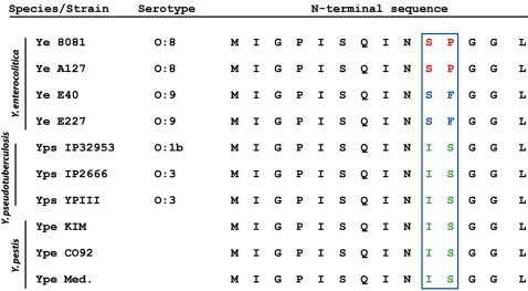
Numerous microbial pathogens modulate or interfere with cell death pathways in cultured cells. However, the precise role of host cell death during in vivo infection remains poorly understood. Macrophages infected by pathogenic species of Yersinia typically undergo an apoptotic cell death. This is due to the activity of a Type III secreted effector protein, designated YopJ in Y. pseudotuberculosis and Y. pestis, and YopP in the closely related Y. enterocolitica. It has recently been reported that Y. enterocolitica YopP shows intrinsically greater capacity for being secreted than Y. pestis YopJ, and that this correlates with enhanced cytotoxicity observed for high virulence serotypes of Y. enterocolitica. The enzymatic activity and secretory capacity of YopP from different Y. enterocolitica serotypes have been shown to be variable. However, the underlying basis for differential secretion of YopJ/YopP, and whether reduced secretion of YopJ by Y. pestis plays a role in pathogenesis during in vivo infection, is not currently known. It has also been reported that similar to macrophages, Y. enterocolitica infection of dendritic cells leads to YopP-dependent cell death. We demonstrate here that in contrast to Y. enterocolitica, Y. pseudotuberculosis infection of bone marrow-derived dendritic cells does not lead to increased cell death. However, death of Y. pseudotuberculosis-infected dendritic cells is enhanced by ectopic expression of YopP in place of YopJ. We further show that polymorphisms at the N-terminus of the YopP/YopJ proteins are responsible for their differential secretion, translocation, and consequent cytotoxicity. Mutation of two amino acids in YopJ markedly enhanced both translocation and cytotoxicity. Surprisingly, expression of YopP or a hypersecreted mutant of YopJ in Y. pseudotuberculosis resulted in its attenuation in oral mouse infection. Complete absence of YopJ also resulted in attenuation of virulence, in accordance with previous observations. These findings suggest that control of cytotoxicity is an important virulence property for Y. pseudotuberculosis, and that intermediate levels of YopJ-mediated cytotoxicity are necessary for maximal systemic virulence of this bacterial pathogen.


Conflict of interest statement

The authors have declared that no competing interests exist.

Figures

Figure 1
Figure 1. Y. enterocolitica YopP induces apoptosis more potently than Y. pseudotuberculosis YopJ in dendritic cells.
(A) PARP cleavage was measured by immunoblotting of macrophage and (B) dendritic cell lysates after infection with indicated Y. pseudotuberculosis (Yp) and Y. enterocolitica (Ye) strains at indicated multiplicity of infection (MOI). P- indicates bacteria lacking the virulence plasmid. Immunoblots are representative of 3 independently infected preparations of cells. (C) NucView™ 488 Caspase-3 fluorescent substrate labeling of dendritic cells infected at an MOI of 5 with indicated bacterial strains.
Figure 2
Figure 2. YopP inhibits MAPK activation to a greater extent than YopJ.
(A) Cell lysates from dendritic cells infected for indicated number of minutes with wild type Y. pseudotuberculosis or enterocolitica (Yp or Ye, respectively) or their virulence plasmid-deficient counterparts (YpP- or YeP-) were probed for phospho-p38 and total p38. (B) Identical cell lysates as in (A) were probed for phospho-SAPK/JNK and total SAPK/JNK. (C) Cell lysates from dendritic cells infected with Yp ΔyopJ expressing either YopP or YopJ as indicated were probed for phospho- and total-MAPK proteins as in (A) and (B). All immunoblots are representative of three independently performed experiments. (D) Flow cytometry analysis of annexinV-positive DCs treated with vehicle (DMSO) or the p38 inhibitor SB202190 immediately prior to being left untreated, treated with LPS, or infected with Yp, YpP-, Ye, or YeP-. Data are represented as bar graphs of the mean of triplicate samples, and are representative of three independently performed experiments.
Figure 3
Figure 3. Extent of YopJ/YopP secretion correlates with degree of dendritic cell death following Yersinia infection.
(A) TCA-precipitated supernatants from indicated bacterial strains analyzed by SDS-PAGE and Coomassie blue staining. (B) Cell lysates from HeLa cells infected with yopJ mutant Y. pseudotuberculosis expressing indicated GSK fusion proteins analyzed by Western blotting for levels of translocation of GSK fusion protein (anti-phospho-Ser9-GSK) and for levels of total GSK fusion protein (anti-GSK3β aa1-13). White arrow indicates phospho-GSK fusion protein, black arrow indicates total GSK-tagged fusion protein. Grey arrows indicate endogenous phospho-GSK and GSK. (C) Percentage of bone marrow derived macrophages that stain positively with annexinV/propidium iodide (PI) 8 hrs post-infection at MOI of 20 with indicated bacterial strains. Dotted line indicates background staining of uninfected cells. (D) Percentage of bone marrow derived dendritic cells that stain positively with PI 16–18 hrs post-infection at MOI of 20 with indicated bacterial strains. Graphs are representative of 3 independently infected preparations of cells, performed in triplicate. p values obtained by Student's unpaired two-tailed t-test.
Figure 4
Figure 4. N-terminal sequence polymorphisms determine differential secretion of YopP/YopJ proteins.
N-terminal 14 amino acids of YopP and YopJ from various Yersinia strains and serotypes. O∶8 serotypes of Y. enterocolitica possess an SP sequence at positions 10 and 11, whereas Y. pseudotuberculosis and Y. pestis possess the IS sequence. O∶9 serotypes of Y. enterocolitica also possess a polymorphism at amino acid 11, which likely accounts for the reduced secretion of YopP by O∶9 serotypes. Ye – Y. enterocolitica, Ype – Y. pestis, Yps – Y. pseudotuberculosis. Ype Med. – Y. pestis, biovar Medievalis.
Figure 5
Figure 5. YopP-expressing Y. pseudotuberculosis causes enhanced killing of cells within lymphoid tissue during mouse infection.
(A) TUNEL staining analyzed by flow cytometry of mesenteric lymph node cells isolated day 4 post-infection from mice given PBS as controls or infected with yopJ mutant Y. pseudotuberculosis expressing YopJ or YopP. CD11b and CD11c populations represent primarily macrophage and dendritic cell populations, respectively. Data are representative of six mice per group for infected mice and three mice for PBS controls. Percentage of double positive cells is indicated in each dot plot. Bar graphs represent mean percentage of double positive cells for six infected mice for each bacterial strain or three PBS control treated mice. (B) Spleen sections from mice infected with YopJ- or YopP- expressing bacteria as in (A), isolated on Day 4 post-infection and stained with TUNEL reagent and DAPI. Scale bar = 40 µM. (C) Enumeration of TUNEL+ nuclei from spleen, mesenteric lymph node, and Peyer's patch sections isolated four days post-infection with YopJ- (light grey bars) or YopP- (dark grey bars) expressing yopJ mutant Y. pseudotuberculosis. Data are averages of at least 10 random fields per mouse per tissue averaged from three mice per group. Similar results were obtained for two independent infections. p values were calculated using the unpaired Student's t-test.
Figure 6
Figure 6. YopP-expressing Y. pseudotuberculosis is attenuated and induces lower levels of serum cytokine production during mouse infection.
(A) Bacterial CFUs present in tissues of mice infected with Yp ΔyopJ containing either empty vector (white triangles) or expressing YopJ (grey squares) or YopP (grey circles) at days 3, 5, and 7 after oral infection. Mice infected with YopJ-expressing Yp ΔyopJ were moribund or dead from overwhelming infection at the end of day 6. (B) Serum cytokine levels of infected mice from (A) collected on days 3 (white bars), 5 (light grey bars), and 7 (dark grey bars). (C) Survival curve of mice infected with YopJ- (grey squares) or YopP- (grey circles) expressing Yp ΔyopJ. p values for bacterial cfu and serum cytokine levels were calculated using unpaired 2-tailed Student's t-Test. p value for mouse survival curves was calculated using log-rank test.

References

    1. Monack D, Falkow S. Apoptosis as a common bacterial virulence strategy. Int J Med Microbiol. 2000;290:7–13. - PubMed
    1. Fu Y, Galan JE. A Salmonella protein antagonizes Rac-1 and Cdc42 to mediate host-cell recovery after bacterial invasion. Nature. 1999;401:293–297. - PubMed
    1. Lin SL, Le TX, Cowen DS. SptP, a Salmonella typhimurium type III-secreted protein, inhibits the mitogen-activated protein kinase pathway by inhibiting Raf activation. Cell Microbiol. 2003;5:267–275. - PubMed
    1. Glomski IJ, Decatur AL, Portnoy DA. Listeria monocytogenes mutants that fail to compartmentalize listerolysin O activity are cytotoxic, avirulent, and unable to evade host extracellular defenses. Infect Immun. 2003;71:6754–6765. - PMC - PubMed
    1. Wren BW. The yersiniae–a model genus to study the rapid evolution of bacterial pathogens. Nat Rev Microbiol. 2003;1:55–64. - PubMed

Publication types

MeSH terms