. 2008 May 16;4(5):e1000065.
doi: 10.1371/journal.ppat.1000065.

Targeting of pseudorabies virus structural proteins to axons requires association of the viral Us9 protein with lipid rafts

Affiliations

Targeting of pseudorabies virus structural proteins to axons requires association of the viral Us9 protein with lipid rafts

Mathew G Lyman et al. PLoS Pathog. .

Abstract

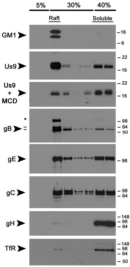
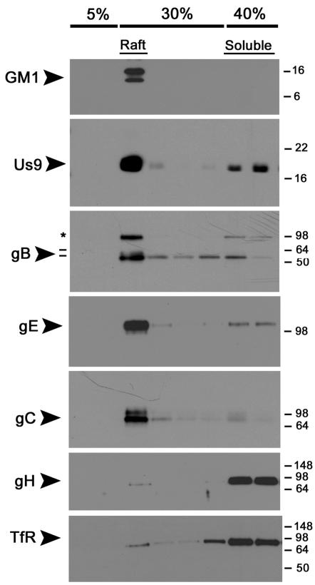
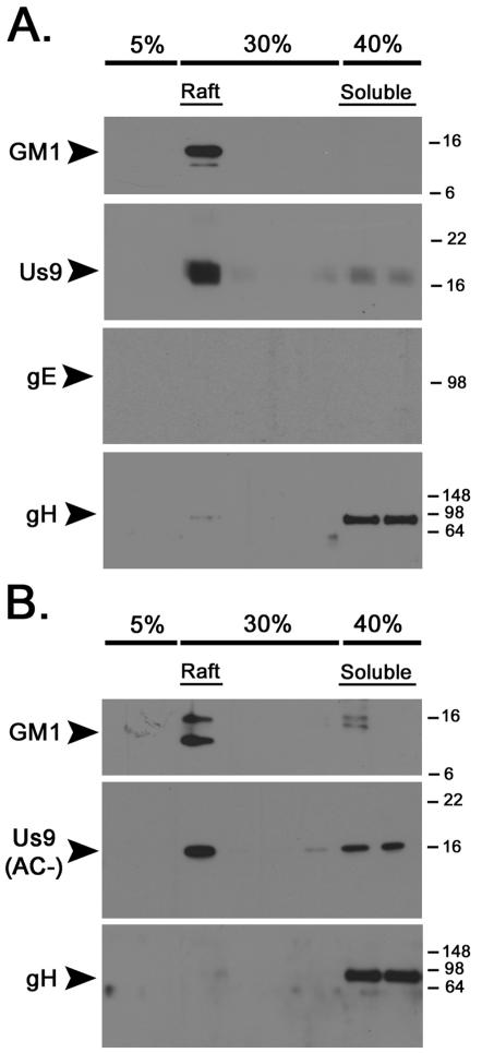
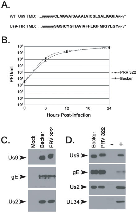
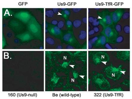
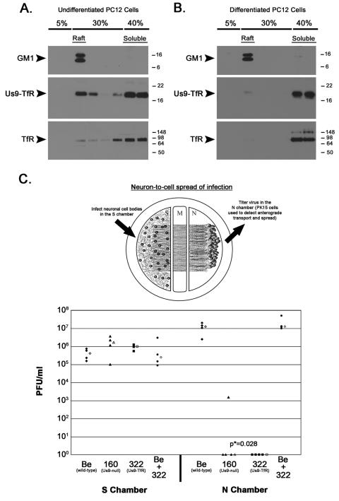
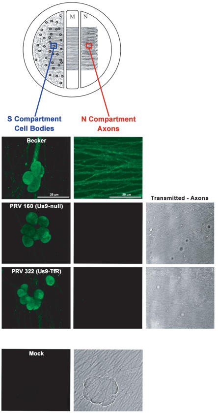
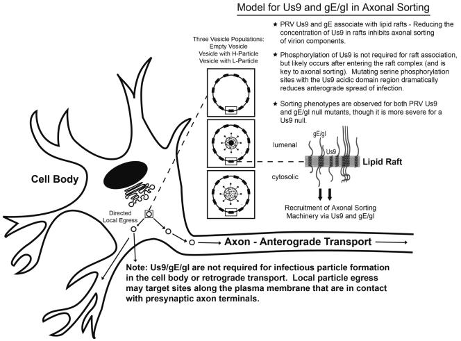
The pseudorabies virus (PRV) Us9 protein plays a central role in targeting viral capsids and glycoproteins to axons of dissociated sympathetic neurons. As a result, Us9 null mutants are defective in anterograde transmission of infection in vivo. However, it is unclear how Us9 promotes axonal sorting of so many viral proteins. It is known that the glycoproteins gB, gC, gD and gE are associated with lipid raft microdomains on the surface of infected swine kidney cells and monocytes, and are directed into the axon in a Us9-dependent manner. In this report, we determined that Us9 is associated with lipid rafts, and that this association is critical to Us9-mediated sorting of viral structural proteins. We used infected non-polarized and polarized PC12 cells, a rat pheochromocytoma cell line that acquires many of the characteristics of sympathetic neurons in the presence of nerve growth factor (NGF). In these cells, Us9 is highly enriched in detergent-resistant membranes (DRMs). Moreover, reducing the affinity of Us9 for lipid rafts inhibited anterograde transmission of infection from sympathetic neurons to epithelial cells in vitro. We conclude that association of Us9 with lipid rafts is key for efficient targeting of structural proteins to axons and, as a consequence, for directional spread of PRV from pre-synaptic to post-synaptic neurons and cells of the mammalian nervous system.


Conflict of interest statement

The authors have declared that no competing interests exist.

Figures

Figure 1
Figure 1. Live-cell imaging of GFP-tagged capsid viruses in differentiated PC12 cells.
Cells were grown on glass-bottom MatTek dishes coated with poly-DL-ornithine and natural mouse laminin prior to imaging on a Leica SP5 confocal microscope. Each frame of the movie, a 2D projection representing a stack of 15 images that are 0.5 µm apart, contains a scale bar (in microns) and timestamp from the movie sequence. (A) A plate of differentiated PC12 cells was infected with PRV GS443 (green capsid) at a low MOI (0.1) and imaged at 12 hours post-infection (hpi). Numerous green capsid puncta were observed in neurites moving in the anterograde direction, i.e. away from the cell body (see Video S1 in supplemental material). This cell was chosen because it had several axon projections to emphasize the sorting phenotypes. Most differentiated PC12 cells have fewer axon projections (1–3). (B) Differentiated PC12 cells were infected with PRV 368 (green capsid, Us9-null) at low MOI and imaged at 12 hpi. The cell body shows robust green fluorescence, but virus capsids were not observed in neurites emanating from the cell body (see Video S2). (C) DIC image showing three neurites extending from the PRV 368-infected cell body (highlighted by the white arrowheads). (D) A differentiated PC12 infected with PRV 368 for 12 hours. No capsids are observed moving in the anterograde direction in two neurites emanating from the cell body (white arrowheads, see Video S3). Note that capsids are not present beyond the proximal segment of the axon. (E and F) Though no green capsid puncta are moving in the anterograde direction (white arrowheads), capsids can be observed moving in a transneuronal, retrograde manner from the infected PC12 cell (red arrowheads, see Video S3) to an uninfected cell above it (panel F, see Video S4). Despite an abundance of moving capsid puncta within the cell body, no other egress events are visible. The brightness in panel F has been increased to better visualize green capsid puncta moving into the uninfected cell body (highlighted by red arrowheads). A white asterisk denotes the accumulation of capsid puncta in the cell body of the uninfected cell.
Figure 2
Figure 2. PRV Us9 and gB are highly-enriched in DRMs of non-polarized PC12 cells, whereas gH is not.
Non-differentiated PC12 Cells were infected with PRV Becker for 12 hours, and then lysed with cold 1% Triton X-100. Lysates were separated on a discontinuous Optiprep™density gradient, and 10 fractions were collected from the top to the bottom of the tube (1 ml each). Samples 2–9 were subjected to SDS-PAGE, and Western blotting analysis was performed using biotinylated cholera toxin B subunit (for GM1) and antibodies to PRV Us9, gB, gE, gC, gH and transferrin receptor (TfR). To test the effect of cholesterol depletion on Us9 association with DRMs, infected PC12 cells were incubated with 20 mM methyl-cyclodextrin (MCD) at 37°C for 45 minutes prior to lysis with cold detergent. The preprocessed form of gB (*) is labeled along with the 69 kDa and 58 kDa (−) subunits.
Figure 3
Figure 3. Differentiation of PC12 cells with nerve growth factor (NGF) increases the association of PRV gE and gC with DRMs, but gH remains soluble.
PC12 cells were differentiated in low serum conditions in the presence of NGF for 12 days. Cells were infected with PRV Becker for 12 hours, and then lysed with cold 1% Triton X-100. Lysates were separated on a discontinuous Optiprep™ density gradient, and analyzed by SDS-PAGE. Western blotting was performed using biotinylated cholera toxin B subunit (for GM1) and antibodies to PRV Us9, gB, gE, gC, gH and transferrin receptor (TfR). The preprocessed form of gB (*) is labeled along with the 69 kDa and 58 kDa (−) subunits.
Figure 4
Figure 4. The raft association of Us9 is not dependent on the presence of gE or the Us9 acidic cluster motif.
Non-differentiated PC12 Cells were infected with PRV 99 (A) or PRV 162 (B) for 12 hours. Cells were subsequently lysed with cold 1% Triton X-100, and separated on a discontinuous Optiprep™ density gradient. Samples were subjected to SDS-PAGE, and the presence of GM1, Us9, gE, and gH was assessed by Western blot analysis.
Figure 5
Figure 5. Characterization of PRV 322 (Us9-TfR).
(A) The amino acid alignment of the C-terminal portion of wild-type Us9 and Us9 with the transferrin receptor transmembrane domain (Us9-TfR). The transmembrane domain sequences are denoted in large, bold letters. (B) Single step growth analysis on Becker and PRV 322 in PK15 cells. (C) The steady-state expression of Us9, gE, and Us2 in PK15 cells infected with Becker and PRV 322 (6 hpi). (D) Incorporation of Us9, Us9-TfR, gE, and Us2 into purified mature virions, but not UL34. Mock and Becker infected lysates serve as the negative (−) and positive (+) controls, respectively.
Figure 6
Figure 6. Localization of Us9-TfR in the absence and presence of infection.
(A) PK15 cells were transfected with plasmids expressing GFP, Us9-GFP, and Us9-TfR-GFP. Cells were fixed with 4% paraformaldehyde at 24 hours post-transfection, and the nuclei stained with Hoechst 33342 (blue). Direct fluorescence was visualized using an inverted epifluorescence microscope using the appropriate excitation and emission filters. The arrowheads highlight the perinuclear, steady-state accumulation of Us9. (B) PK15 cells were infected with PRV 160 (Us9-null), Becker (wild-type), or PRV 322 (Us9-TfR) for 6 hours. Cells were fixed and stained with Us9 antiserum, and visualized on a Leica SP5 confocal microscope. Arrowheads denote Us9 accumulations adjacent to the cell nucleus (N).
Figure 7
Figure 7. Raft association of Us9 is critical to anterograde spread of infection in vitro.
(A) Non-differentiated and (B) differentiated PC12 Cells were infected with PRV 322 for 12 hours, and then lysed with cold 1% Triton X-100. Lysates were separated on a discontinuous Optiprep™ density gradient, and analyzed by SDS-PAGE. Western blot analysis was performed using biotinylated cholera toxin B subunit (for detection of GM1), and antiserum specific for Us9 and TfR. (C) Trichamber diagram illustrating the system used to measure PRV anterograde spread of infection from neurons to PK15 cells. SCG neurons were plated in the S chamber and allowed to extend neurites into the N chamber. The neurites were guided into the N compartment by a series of grooves. A monolayer of indicator PK15 cells were then plated on top of the axon termini in the N chamber. Cell bodies in the S chamber were infected, virus particles sorted into axons in a Us9-dependent manner, and these particles subsequently infected the PK15 cells that amplified the infection. Cultured neurons in the S chamber were infected at a high MOI with Becker (wild-type), PRV 160 (Us9-null), PRV 322 (Us9-TfR), or both Becker and PRV 322. Four chambers were used for each type of infection (closed symbols). At 24 hpi, medium and infected cells were harvested together from either the S or N chambers. Total plaque-forming units (PFU)/ml were determined for each chamber. The median value for the four samples is denoted by the offset open symbol. The P value (p*) was determined using the Wilcoxon two-sample test.
Figure 8
Figure 8. PRV 322 (Us9-TfR) is defective in axonal sorting of virus structural proteins.
Trichamber diagram illustrating the system used to visualize PRV antigens in the cell bodies and axons of infected neurons. The blue box illustrates the site within the S compartment were cell bodies were imaged. The red box indicates the site were axons were imaged in the N compartment. SCG neurons were plated in the S chamber and allowed to extend neurites into the N chamber (through the M compartment). The neurites were guided into the N compartment by a series of grooves. Two weeks post-plating, cell bodies in the S chamber were infected at a high MOI with Becker (wild-type), PRV 160 (Us9-null) or PRV 322 (Us9-TfR). At 16 h postinfection, samples were fixed and labeled with PRV-specific polyclonal antiserum (Rb134) that recognizes virus glycoprotein and virus capsid proteins. All infected cell bodies within the S compartment stained for viral structural proteins (first column). Mock-infected cells did not label with the Rb134 antibody. Becker-infected axons stained heavily for PRV antigen (second column), though axons from PRV 160 and PRV 322 infected cell bodies were devoid of viral glycoprotein and capsid proteins (though an extensive network of axons was visible by transmitted brightfield illumination).
Figure 9
Figure 9. The role of Us9, gE/gI, and lipid rafts in anterograde sorting.
Us9 and gE/gI enter the secretory pathway and associate with lipid rafts in the trans-Golgi network (TGN), the site of viral assembly. The presence of Us9 and gE/gI in lipid rafts, and subsequent Us9 phosphorylation, would recruit axonal sorting machinery to the cytoplasmic face of a small number of viral assembly complexes in the TGN, i.e. vesicles with viral membrane proteins only (empty vesicle), those containing mature virus particles (H-particle), or L-particles. A small number of assembly complexes would then bind a sorting adaptor protein(s) and go to axons; the majority of assembly complexes would target sites along the plasma membrane that are in contact with presynaptic axon terminals. Random egress is rare.

References

    1. Brown A. Axonal transport of membranous and nonmembranous cargoes: a unified perspective. J Cell Biol. 2003;160:817–821. - PMC - PubMed
    1. Dotti CG, Simons K. Polarized sorting of viral glycoproteins to the axon and dendrites of hippocampal neurons in culture. Cell. 1990;62:63–72. - PubMed
    1. Edidin M. The state of lipid rafts: from model membranes to cells. Annu Rev Biophys Biomol Struct. 2003;32:257–283. - PubMed
    1. Ledesma MD, Brugger B, Bunning C, Wieland FT, Dotti CG. Maturation of the axonal plasma membrane requires upregulation of sphingomyelin synthesis and formation of protein-lipid complexes. Embo J. 1999;18:1761–1771. - PMC - PubMed
    1. Rajendran L, Simons K. Lipid rafts and membrane dynamics. J Cell Sci. 2005;118:1099–1102. - PubMed

Publication types

MeSH terms