. 2008 May 16;4(5):e1000073.
doi: 10.1371/journal.pgen.1000073.

HOXA13 Is essential for placental vascular patterning and labyrinth endothelial specification

Affiliations

HOXA13 Is essential for placental vascular patterning and labyrinth endothelial specification

Carley A E Shaut et al. PLoS Genet. .

Abstract

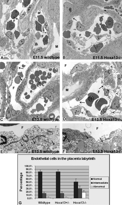
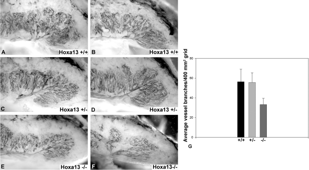
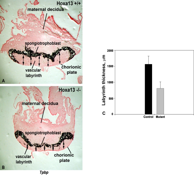
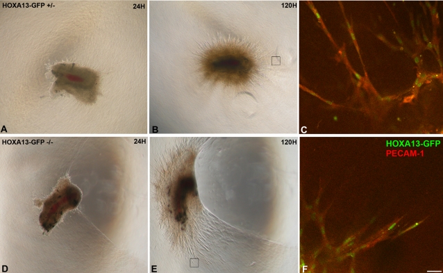
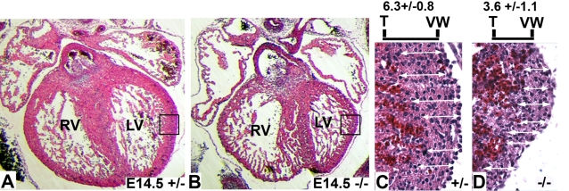
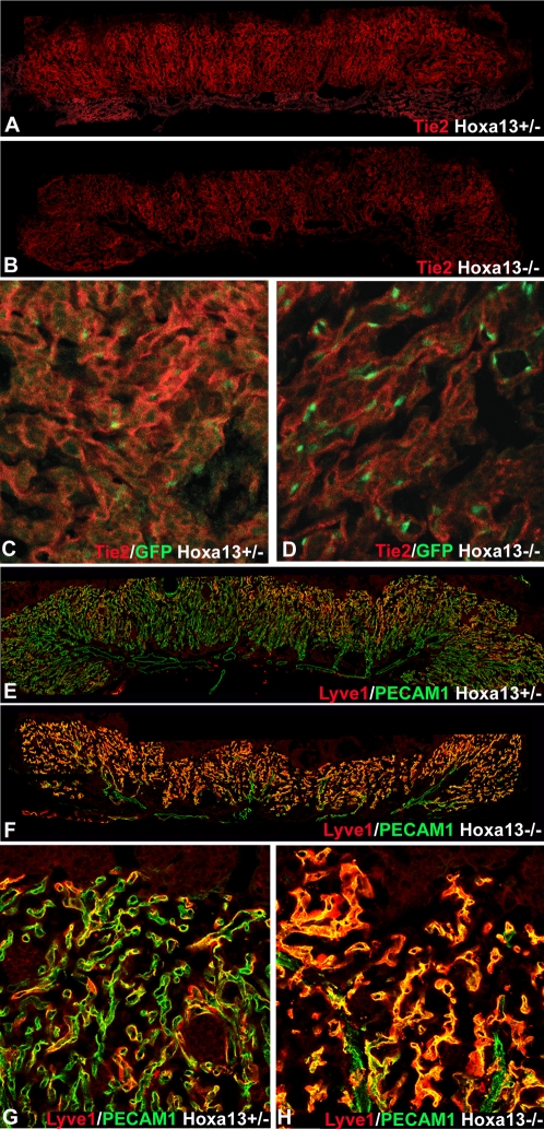
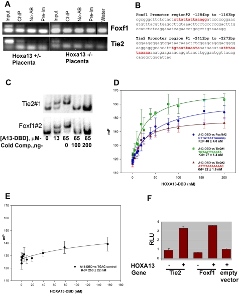
In eutherian mammals, embryonic growth and survival is dependent on the formation of the placenta, an organ that facilitates the efficient exchange of oxygen, nutrients, and metabolic waste between the maternal and fetal blood supplies. Key to the placenta's function is the formation of its vascular labyrinth, a series of finely branched vessels whose molecular ontogeny remains largely undefined. In this report, we demonstrate that HOXA13 plays an essential role in labyrinth vessel formation. In the absence of HOXA13 function, placental endothelial cell morphology is altered, causing a loss in vessel wall integrity, edema of the embryonic blood vessels, and mid-gestational lethality. Microarray analysis of wild-type and mutant placentas revealed significant changes in endothelial gene expression profiles. Notably, pro-vascular genes, including Tie2 and Foxf1, exhibited reduced expression in the mutant endothelia, which also exhibited elevated expression of genes normally expressed in lymphatic or sinusoidal endothelia. ChIP analysis of HOXA13-DNA complexes in the placenta confirmed that HOXA13 binds the Tie2 and Foxf1 promoters in vivo. In vitro, HOXA13 binds sequences present in the Tie2 and Foxf1 promoters with high affinity (K(d) = 27-42 nM) and HOXA13 can use these bound promoter regions to direct gene expression. Taken together, these findings demonstrate that HOXA13 directly regulates Tie2 and Foxf1 in the placental labyrinth endothelia, providing a functional explanation for the mid-gestational lethality exhibited by Hoxa13 mutant embryos as well as a novel transcriptional program necessary for the specification of the labyrinth vascular endothelia.


Conflict of interest statement

The authors have declared that no competing interests exist.

Figures

Figure 1
Figure 1. Early expression of Hoxa13 in the allantois and its derivatives.
(A, B) HOXA13 is expressed in the umbilical arteries which partially stenose (white arrow) in Hoxa13 homozygous mutants. (C) Fluorescent and bright field image of an E7.75 embryo expressing HOXA13-GFP. AL = allantois, CC = cardiac crescent. (D) HOXA13-GFP is expressed throughout the allantois at E8.5. Red signal = detection of the HOXA13-GFP fusion protein using a GFP antibody (denoted as α-GFP). Green signal indicates detection of the endogenous HOXA13-GFP fusion protein. Yellow signal (Merged) indicates the co-localization of the detected HOXA13-GFP protein and the α-GFP immuno-positive cells, confirming the detected green fluorescence to be derived from the mutant HOXA13-GFP fusion protein throughout the allantois. AL = allantois; EM = embryo proper. (E, F) Cryosection of an E9.5 placenta. Green signal indicates detection of the HOXA13-GFP fusion protein in the developing labyrinth region; red signal indicates detection of the HOXA13-GFP fusion protein using a GFP antibody (denoted as α-GFP). Note the absence of HOXA13-GFP expression in the chorionic ectoderm (CE). Arrows denote sites of microvessel genesis. Dashed line represents chorionic plate. AS = allantoic stalk. (G) Sagittal section of an E9.5 embryo and developing placenta. Note that HOXA13-GFP expression is maintained in the allantoic stalk as it contributes to the developing allantoic vessels (arrows) as well as the developing chorionic plate vessels (asterisks). Dashed line denotes the developing chorionic plate. CE = chorionic ectoderm, EM = embryo proper, AS = allantoic stalk. (H) At E10.5, HOXA13 expression is maintained in the developing labyrinth (LAB), whereas little or no expression is detected in the chorionic ectoderm (CE). Bars are 25 µm.
Figure 2
Figure 2. Sites of HOXA13 expression during placental labyrinth development.
(A) HOXA13 is expressed in the endothelial progenitors in the E8.0 allantois. (B) HOXA13 expression is maintained in the endothelial progenitors during chorioallantoic fusion at E8.5. (C) Between E8.75 and E9.5, the HOXA13-expressing endothelial progenitors contribute to the developing feto-placental vessels, which mature to form the umbilical artery (UA), chorionic plate vessels, and the vessels contributing to the placental labyrinth (D). HOXA13 is not expressed in the developing umbilical vein (UV).
Figure 3
Figure 3. HOXA13 is expressed in the placental labyrinth vascular endothelia.
(A, B) Analysis of HOXA13 expression (green) in heterozygous control and homozygous mutant placentas at E12.5 reveals extensive expression throughout the vascular labyrinth. (C) HOXA13 is co-expressed with PECAM-1 in the labyrinth endothelia, which exhibit an elongated morphology in heterozygous controls (arrowheads). (D) E12.5 homozygous mutant labyrinth endothelia also express HOXA13 (green signal) and PECAM-1 (red signal), but maintain a rounded morphology (arrowheads). (E, F) Analysis of labyrinth vessels at E10.5 reveals co-localization of HOXA13 (green signal) and PECAM-1 (red signal) to the undifferentiated endothelia (arrowheads), which do not exhibit an elongated vascular morphology. Bar is 10 µm for (C–F).
Figure 4
Figure 4. Endothelial cell morphology is affected in the placental labyrinth of Hoxa13 homozygous mutants.
(A, B) Transmission electron microscopy (TEM) reveals the initial elongation of the wild-type EC in the developing labyrinth vessels, whereas homozygous mutant littermates (B) exhibit rounded endothelia with attenuated cell bodies. Asterisks denote the endothelial cells; arrows depict the EC bodies in wild-type and mutant vessels. (C, D) Wild-type controls exhibit a mature elongated endothelial cell morphology by E13.5 (arrows), whereas homozygous mutant littermates (D) exhibit a severe loss in the elongated morphology (arrows), causing edema in the surrounding placental tissues (arrowheads). F = fetal vessel lumen; M = maternal space. (E, F) No differences in basement membrane (arrows) ultrastructure were detected in the labyrinth vessels between wild-type and homozygous mutant embryos, confirming that the loss of HOXA13 function is directly affecting endothelial morphology and function. F = fetal vessel lumen; N = endothelial cell nucleus. (G) Quantitative analysis of multiple labyrinth vessel TEM micrographs revealed that nearly 50% of the homozygous mutant endothelia exhibited an intermediate or severely abnormal morphology compared to wild-type or heterozygous mutant controls.
Figure 5
Figure 5. Labyrinth vessel branching is reduced in Hoxa13 homozygous mutants.
(A–D) PECAM-1 immunostaining of E13.5 hemisected placentas revealed extensive branching of the primary labyrinth vessel in wild-type and heterozygous mutants, whereas homozygous mutant littermates exhibited poor branching of the primary labyrinth vessels (E, F). (G) Quantitation of the labyrinth vascular branches confirmed that the homozygous mutants labyrinths contained an average of 33 branches per 400 mm2 grid, whereas wild-type and heterozygous mutant controls contained an average of 56 and 55 branches, respectively, in the same unit area. Bars represent the standard deviation of six independent assessments.
Figure 6
Figure 6. The vascular labyrinth region is thinner in Hoxa13 homozygous mutant placentas.
(A), (C) Measurements of the E12.5 labyrinth, which is defined as the region underlying the spongiotrophoblast marker, Tpbp, revealed an average labyrinth thickness of nearly 1,600 µm in Hoxa13 control embryos. (B, C) Parallel analyses of age-matched Hoxa13 homozygous mutants confirmed that the labyrinth thickness is reduced to an average thickness of 800 µm. Arrows represent the sites measured on each labyrinth section from the spongiotrophoblast to the chorionic plate to determine labyrinth thickness using the NIH Image J software. Error bars represent the standard deviation of 16 different labyrinth sections measured at 5 independent points for each sample.
Figure 7
Figure 7. Analysis of endothelial cell migration and neovascularization in cultured placental primary arteries.
(A, B), (D, E) Arterial sections from heterozygous control and homozygous mutants exhibit robust neovascularization and microvessel formation in vitro. Black boxes denote the sites examined by confocal microscopy to visualize migrating endothelia participating in microvessel formation. (C), (F) The loss of HOXA13 function does not affect the migration and contribution of HOXA13-expressing endothelia (green nuclei) to the developing microvessels. Note that the migrating endothelia co-express PECAM-1 (red signal). Bar is 25 µm.
Figure 8
Figure 8. Analysis of ventricular wall thickness in Hoxa13 mutants.
(A, B) Analysis of hematoxylin and eosin–stained sections (7 µm) from heterozygous control and homozygous mutant hearts at E14.5 embryos reveal qualitatively thicker ventricular walls in the heterozygous control embryos. RV = right ventricle; LV = left ventricle. Boxes represent the enlarged regions shown in (C) and (D). (C, D) Quantitative analysis of the left ventricular wall thicknesses revealed homozygous mutants possess an average of 3.6 (±1.1) cells per linear assessment versus 6.3 (±0.8) cells in heterozygous controls, confirming that the E14.5 Hoxa13 homozygous mutants possess a thinner left ventricular wall compared to age-matched heterozygous controls. White arrows depict the sites assessed for ventricular wall thickness using the hematoxylin-stained nuclei to determine cell number. The measurements were taken from the trabecular wall (T) to the outer ventricular wall (VW) for each assessment.
Figure 9
Figure 9. TIE-2 and LYVE-1 are co-expressed with HOXA13 in the labyrinth vascular endothelia and exhibit altered expression in E13.5 homozygous mutants.
(A, B) Immunohistochemical analysis of TIE2 expression (red signal) in the placental labyrinth reveals reduced levels in E13.5 Hoxa13 homozygous mutants compared to heterozygous controls. (C, D) Higher magnification image of the placental labyrinth confirms that TIE2 (red signal) is co-expressed with HOXA13 (green nuclear signal) in the labyrinth vascular endothelia. (E, F) Immunohistochemical analysis of LYVE-1 expression (red signal) in the placental labyrinths reveals elevated levels of LYVE-1 in E13.5 Hoxa13 homozygous mutants compared to heterozygous controls. (G, H) Analysis of LYVE-1 expression at higher magnification (red signal) confirms that the PECAM-1–positive endothelial cells (green signal) co-express LYVE-1 and that LYVE-1 expression is elevated in the labyrinth vasculature of Hoxa13 homozygous mutants.
Figure 10
Figure 10. HOXA13 binds to gene-regulatory regions in Tie2 and Foxf1 to facilitate gene expression.
(A). Left panel: Chromatin immunoprecipitation (ChIP) of heterozygous control placental chromatin using a HOXA13 antibody identified DNA sequences present in the Tie2 and Foxf1 promoter regions and confirms that HOXA13 associates with these sequences in vivo. Right panel: Parallel assays using Hoxa13 homozygous mutant placentas failed to enrich for the same DNA regions, suggesting that HOXA13's DNA binding function, which is absent in the homozygous mutant protein, is necessary for in vivo association at the Tie2 and Foxf1 loci. Input indicates positive control confirming the presence of the Tie2 and Foxf1 DNA elements in both heterozygous and homozygous mutant chromatin samples. ChIP = + HOXA13 antibody; No-AB = no antibody negative control; Pre-Im = IgG control rabbit pre-immune sera; Water = no DNA PCR control (B). Sequence analysis revealed HOXA13 binding sites (red text) in the ChIP-positive PCR amplification products for both Foxf1 and Tie2. (C) EMSA analysis of the ChIP-positive PCR amplification products confirms HOXA13 can bind these sites in a concentration-dependent manner. A13-DBD = HOXA13 DNA binding domain peptide; Cold Comp.  = the Tie2 or Foxf1 binding sites lacking radioactive labeling. (D) The HOXA13 DNA binding domain binds the DNA sequences present in the ChIP-positive region with high affinity exhibiting a Kd of 48±4 nM for the Foxf1 region and 27±1.4 nM and 22±1.6 nM for the two sequences present in Tie2. Error bars denote the standard error for the averaged millipolarization values at each HOXA13-DBD concentration. The DNA sequences of the Tie2 or Foxf1 promoter regions used in the assay are denoted by the color text. (E) Conversion of the Tie2 or Foxf1 binding sites to the sequence TGAC ablates the affinity of the HOXA13 DNA binding domain for these gene-specific promoter sequences. Error bars denote the standard error for the averaged millipolarization values at each HOXA13-DBD concentration. (F) In vitro assessment of the 140 bp Tie2 and 121 bp Foxf1 ChIP fragments confirms that HOXA13 can use these minimal sites to direct gene expression, confirming their capacity to function as gene-regulatory elements. Values represent average detected luciferase activity after normalization for transfection with a Renilla luciferase reporter±standard error.

References

    1. Rossant J, Cross JC. Placental development: lessons from mouse mutants. Nat Rev Genet. 2001;2:538–548. - PubMed
    1. Breier G. Angiogenesis in embryonic development–a review. Placenta. 2000;21(Suppl A):S11–15. - PubMed
    1. Cross JC, Hemberger M, Lu Y, Nozaki T, Whiteley K, et al. Trophoblast functions, angiogenesis and remodeling of the maternal vasculature in the placenta. Mol Cell Endocrinol. 2002;187:207–212. - PubMed
    1. Downs KM. The murine allantois. Curr Top Dev Biol. 1998;39:1–33. - PubMed
    1. Downs KM. Early placental ontogeny in the mouse. Placenta. 2002;23:116–131. - PubMed

Publication types

MeSH terms