Review
. 2008 Jun;291(6):653-60.
doi: 10.1002/ar.20664.

Functional anatomy of normal bile ducts

Affiliations
Review

Functional anatomy of normal bile ducts

Mario Strazzabosco et al. Anat Rec (Hoboken). 2008 Jun.

Abstract

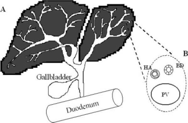
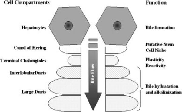
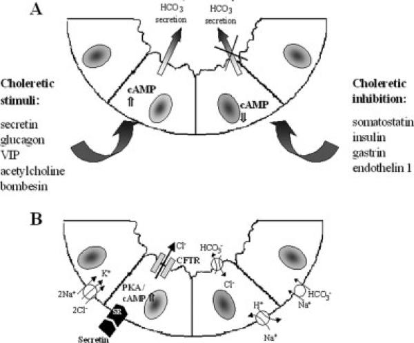
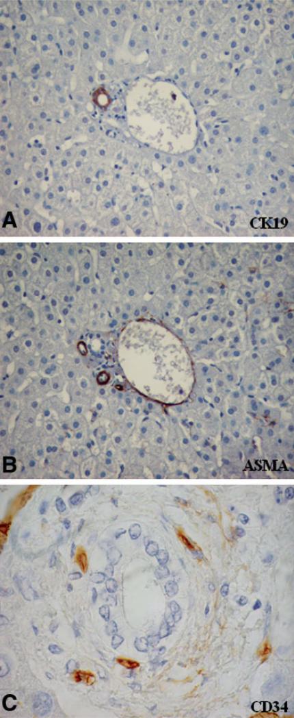
The biliary tree is a complex network of conduits that begins with the canals of Hering and progressively merges into a system of interlobular, septal, and major ducts which then coalesce to form the extrahepatic bile ducts, which finally deliver bile to the gallbladder and to the intestine. The biliary epithelium shows a morphological heterogeneity that is strictly associated with a variety of functions performed at the different levels of the biliary tree. In addition to funneling bile into the intestine, cholangiocytes (the epithelial cells lining the bile ducts) are actively involved in bile production by performing both absorbitive and secretory functions. More recently, other important biological properties restricted to cholangiocytes lining the smaller bile ducts have been outlined, with regard to their plasticity (i.e., the ability to undergo limited phenotypic changes), reactivity (i.e., the ability to participate in the inflammatory reaction to liver damage), and ability to behave as liver progenitor cells. Functional interactions with other branching systems, such as nerve and vascular structures, are crucial in the modulation of the different cholangiocyte functions.


Figures

Fig. 1
Fig. 1
Normal anatomy of biliary epithelium. A: The intrahepatic bile duct epithelium is organized as a three-dimensional branching system of conduits inside the liver, which progressively merge into ducts of increasing size and ultimately deliver bile to the gallbladder and to the duodenum. B: In the liver microarchitecture bile ducts (BD) run in parallel between the lobules with a branch of the portal vein (PV) and of the hepatic artery (HA), giving rise to a close anatomic association known as portal triad.
Fig. 2
Fig. 2
Morphological and functional heterogeneity of biliary epithelium. The biliary tree begins with the canals of Hering, located at the ductular-hepatocellular junction, which are lined in part by hepatocytes and in part by cholangiocytes: they constitute the physiologic link of the biliary tree with the hepatocyte canalicular system, and are the site where a facultative progenitor cell compartment resides. Cells lining the intrahepatic biliary tree have different functional and morphological specializations: the terminal cholangioles (size <15 μm) have some biological properties such as plasticity and reactivity; interlobular (15–100 μm) and large ducts (100 μm to 800 μm) modulates fluidity and alkalinity of the primary hepatocellular bile.
Fig. 3
Fig. 3
Cholangiocyte modifies bile flow and alkalinity by exerting both secretory and absorptive activities. A: Cholangiocyte possesses numerous different molecules located on their apical (luminal) and basolateral domain finely regulated by a balance of prosecretory and antisecretory signals induced by a series of hormones, neuropeptides and neurotrasmitters interacting at the level of adenyl-cyclase. B: In particular, a relevant role in normal cholangiocyte physiology is played by secretin that increases intracellular cAMP levels and consequently activates CFTR through PKA phosphorylation: the consequent Cl efflux into the lumen generates the driving force for HCO3 secretion by AE2 exchanger into bile.
Fig. 4
Fig. 4
Preferential association of the bile ducts with hepatic artery branches in normal portal tract and localization of peribiliary vascular plexus (PBP) around the intrahepatic bile duct. A,B: In the normal portal tract bile ducts (immunoreactive for cytokeratin-19, CK19, A) are closely associated with one or two branches of hepatic artery (immunoreactive for smooth muscle actin, SMA, B): their numerical correlation is considered a good index to measure bile duct preservation or loss. C: Blood is supplied to the bile ducts through a fine system of arterioles and capillaries (recognized by immunoreactivty for CD34, a specific marker of the endothelial phenotype), derived from the hepatic artery, which closely surrounds the cholangiocytes and drains into venules joining the portal vein system. Serial sections of a liver sample with minimal histological changes (nearly normal liver). Original magnifications: ×400 in A,B, ×1,000 in C.
Fig. 5
Fig. 5
Different expression of VEGF, VEGFR-1 and VEGFR-2 on normal bile ducts and reactive ductules. A–D: Normal bile ducts (identified by immunoreactivity for CK19, A) are negative for VEGF (B) and for its receptors VEGFR-1 (C) and VEGFR-2 (D); noteworthy, VEGFR-1 is expressed by endothelial cells lining both portal vein and hepatic artery branches (C). E–H: In contrast, in ductular reaction cholangiocytes (immunostained by CK19, E) show a strong immunoreactivity for VEGF (F), along with VEGFR-1 (G), and VEGFR-2 (H), which appear to be both up-regulated in disease conditions. Liver samples were obtained from a patient with minimal histological changes (A–D) and from a patient with extrahepatic bile duct atresia as example of ductular reaction (E–H). Original magnifications: ×400 in A–H.

Similar articles

Cited by

References

    1. Alvaro D, Alpini G, Jezequel AM, Bassotti C, Francia C, Fraioli F, Romeo R, Marucci L, LeSage G, Glaser SS, Benedetti A. Role and mechanisms of action of acetylcholine in the regulation of cholangiocyte secretory functions. J Clin Invest. 1997;100:1349–1362. - PMC - PubMed
    1. Alvaro D, Mancino MG, Glaser S, Gaudio E, Marzioni M, Francio H, Alpini G. Proliferating cholangiocytes: a neuroendocrine compartment in the diseased liver. Gastroenterology. 2007;132:415–431. - PubMed
    1. Benedetti A, Bassotti C, Rapino K, Marucci L, Jezequel AM. A morphometric study of the epithelium lining the rat intrahepatic biliary tree. J Hepatol. 1996;24:335–342. - PubMed
    1. Caligiuri A, Glaser S, Rodgers RE, Phinizy JL, Robertson W, Papa E, Pinzani M, Alpini G. Endothelin-1 inhibits secretinstimulated ductal secretion by interaction with ETA receptors on large cholangiocytes. Am J Physiol. 1998;275:G835–G846. - PubMed
    1. Cho WK, Boyer JL. Vasoactive intestinal polypeptide is a potent regulator of bile secretion in rat cholangiocytes. Gastroenterology. 1999a;117:420–428. - PubMed

Publication types

MeSH terms

Substances

LinkOut - more resources