Review
. 2008 May 16;3(5):268-81.
doi: 10.1021/cb800037d.

Molecular electron microscopy: state of the art and current challenges

Affiliations
Review

Molecular electron microscopy: state of the art and current challenges

Henning Stahlberg et al. ACS Chem Biol. .

Abstract

The objective of molecular electron microscopy (EM) is to use electron microscopes to visualize the structure of biological molecules. This Review provides a brief overview of the methods used in molecular EM, their respective strengths and successes, and current developments that promise an even more exciting future for molecular EM in the structural investigation of proteins and macromolecular complexes, studied in isolation or in the context of cells and tissues.


Figures

Figure 1
Figure 1
Structure of the AQP0-mediated membrane junction at 1.9 Å resolution obtained by electron crystallography. a) The three water molecules (white arrows) in the water channel of AQP0. b) The two lipid bilayers of the membrane junction with the modeled structures of the lipid molecules. c) Atomic model of an AQP0 subunit with the nine surrounding lipid molecules. Figure adapted from ref (17). Reprinted by permission from Macmillan Publishers Ltd., copyright 2005.
Figure 2
Figure 2
Single-particle EM of the rotavirus inner capsid particle. a) Cryo-EM image of rotavirus inner capsid particles in vitrified ice. The arrows indicate partially damaged particles. b) Overview of the 13-fold averaged viral protein 6 (VP6) trimer at 3.8 Å resolution. The area outlined in red is shown in more detail in panels c and d. c) and d) Density outlined in panel b before (panel c) and after (panel d) 13-fold averaging with the fit crystal structure of VP6 (B. McLain, E. Settembre, R. Bellamy, and S. C. Harrison, unpublished data). Figure adapted from ref (28). Copyright 2008, National Academy of Sciences, U.S.A.
Figure 3
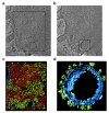
Figure 3
Cryo-electron tomography of a peripheral region of a Dictyostelium cell. a) and b) 60 nm thick slices through the electron tomogram. Scale bar is 200 nm. c) Surface rendering of the volume indicated in (b), showing the actin network (red), membranes (blue), and cytoplasmic macromolecular complexes (green). d) Surface rendering of the volume indicated in (b), showing part of the rough endoplasmatic reticulum with ribosome-like densities (green) decorating the membrane (blue). Figure adapted from ref (34). Copyright 2002. Reprinted with permission from AAAS.
Figure 4
Figure 4
Models of complexes obtained by placing atomic models into single-particle reconstructions. a) Model of an interferon−receptor complex produced by visually placing the atomic models of the subunits into an ∼30 Å density map without computational refinement. Such models only provide information on the approximate spatial relationship between the subunits. Figure adapted from ref (91), Copyright 2008. Reprinted with permission from Elsevier. b) Pseudoatomic model of the transferrin−transferrin receptor complex produced by docking atomic models of the subunits into an ∼8 Å density map with subsequent computational refinement. The transferrin residues interacting with the receptor were confirmed by mutagenesis. Figure adapted from ref (25), Copyright 2004. Reprinted with permission from Elsevier.

References

    1. Wade R. H. (1992) A brief look at imaging and contrast transfer. Ultramicroscopy 46, 145–156.
    1. Scherzer O. (1948) The theoretical resolution limit of the electron microscope. Appl. Phys. 20, 20–29.
    1. Zhu J.; Penczek P. A.; Schröder R.; Frank J. (1997) Three-dimensional reconstruction with contrast transfer function correction from energy-filtered cryoelectron micrographs: procedure and application to the 70S Escherichia coli ribosome. J. Struct. Biol. 118, 197–219. - PubMed
    1. Adrian M.; Dubochet J.; Fuller S. D.; Harris J. R. (1998) Cryo-negative staining. Micron 29, 145–160. - PubMed
    1. Golas M. M.; Sander B.; Will C. L.; Lührmann R.; Stark H. (2003) Molecular architecture of the multiprotein splicing factor SF3b. Science 300, 980–984. - PubMed

Publication types