. 2008 Jul;69(2):402-17.
doi: 10.1111/j.1365-2958.2008.06289.x.

A novel pathway of intercellular signalling in Bacillus subtilis involves a protein with similarity to a component of type III secretion channels

Affiliations

A novel pathway of intercellular signalling in Bacillus subtilis involves a protein with similarity to a component of type III secretion channels

Amy H Camp et al. Mol Microbiol. 2008 Jul.

Abstract

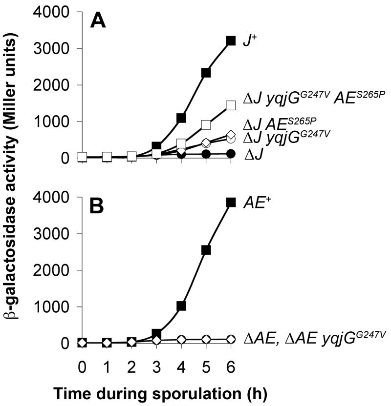
During spore formation in Bacillus subtilis, sigma(E)-directed gene expression in the mother-cell compartment of the sporangium triggers the activation of sigma(G) in the forespore by a pathway of intercellular signalling that is composed of multiple proteins of unknown function. Here, we confirm that the vegetative protein SpoIIIJ, the forespore protein SpoIIQ and eight membrane proteins (SpoIIIAA through SpoIIIAH) produced in the mother cell under the control of sigma(E) are ordinarily required for intercellular signalling. In contrast, an anti-sigma(G) factor previously implicated in the pathway is shown to be dispensable. We also present evidence suggesting that SpoIIIJ is a membrane protein translocase that facilitates the insertion of SpoIIIAE into the membrane. In addition, we report the isolation of a mutation that partially bypasses the requirement for SpoIIIJ and for SpoIIIAA through SpoIIIAG, but not for SpoIIIAH or SpoIIQ, in the activation of sigma(G). We therefore propose that under certain genetic conditions, SpoIIIAH and SpoIIQ can constitute a minimal pathway for the activation of sigma(G). Finally, based on the similarity of SpoIIIAH to a component of type III secretion systems, we speculate that signalling is mediated by a channel that links the mother cell to the forespore.


Figures

Figure 1
Figure 1. σG activation is tightly coupled to σE–directed gene expression independently of sigG transcriptional regulation and the antisigma factors CsfB and SpoIIAB
A. Cartoon depicting the morphological stages of B. subtilis sporulation and the cell-cell signaling pathways controlling σ factor activation in each compartment. Immediately following asymmetric septation, σF becomes active in the smaller forespore compartment and initiates a signal transduction pathway that leads to σE activation in the larger mother cell. Upon the completion of engulfment, σE directs the activation of σG in the forespore by a poorly understood mechanism. Finally, active σG initiates a signal transduction pathway that activates σK in the mother cell. B. σG activity commences during the third hour of sporulation. σG–dependent expression of a translational PsspB-lacZ reporter gene was monitored during sporulation of cells expressing sigG from its normal promoter and at its normal position (solid squares, strain AHB41). C. The timing of σG activation and its dependence on intercellular signaling are maintained in cells expressing PspoIIQ-sigG in place of the endogenous sigG gene. σG–dependent PsspB-lacZ expression during sporulation was monitored in wild type (solid squares), sigE (open circles), Q (open squares), AA-AH (open diamonds), or J (open triangles) mutant cells, all of which expressed PspoIIQ-sigG in place of the endogenous sigG gene (strains AHB350, AHB415, AHB363, AHB411, and AHB365, respectively). D. The timing of σG activation and its dependence on mother cell gene expression are unaffected by the absence of CsfB. The σG–dependent accumulation of β-galactosidase from the PsspB-lacZ reporter gene in wild type (closed squares), csfB (open squares), sigE (closed triangles), and sigE csfB double mutant cells (open triangles) was monitored through sporulation (strains AHB563, AHB580, AHB608, and AHB611, respectively). All strains expressed PspoIIQ-sigG in place of the endogenous sigG gene. A slight increase in the basal levels of σG activity in csfB mutant cells can be observed when the y-axis scale is changed (inset). E. The timing of σG activation and its dependence on mother cell gene expression remains intact in the absence of regulation by the antisigma factors SpoIIAB and CsfB. σG–dependent PsspB-lacZ expression during sporulation was monitored in wild type (closed squares), csfB (open squares), sigE (closed triangles), and sigE csfB double mutant cells (open triangles), all of which expressed PspoIIQ-sigGE156K in place of the endogenous sigG gene (strains AHB565, AHB582, AHB610, and AHB613, respectively). A slight increase in the basal levels of σG activity in csfB mutant cells can be observed when the y-axis scale is changed (inset).
Figure 2
Figure 2. Isolation of J suppressor mutants partially restored for spore formation and σG activation
A. Spore formation is partially restored to J suppressor mutants a-e. Cells were induced to sporulate for 24 hours in DSM medium, and spores were measured as heat resistant colony forming units. With the exception of the parental wild-type strain (WT; PY79), all strains expressed PspoIIQ-sigG in place of endogenous sigG and were deleted for csfB. Cells harbored a wild-type copy of the J gene (J+; AHB480), lacked JJ; AHB434), or lacked J and harbored one of the five indicated suppressors (ΔJ suppressors a-e; AHB442, AHB547, AHB543, AHB438, AHB551, respectively). Error bars indicate standard deviation. B. σG activation is partially restored to J mutant cells harboring suppressors a-e. σG–dependent PsspB-lacZ expression was monitored during sporulation of cells harboring a wild-type copy of the J gene (J+; AHB352), lacking JJ; AHB366), or lacking J and harboring one of the five indicated suppressors (ΔJ suppressors a-e; AHB419, AHB422, AHB420, AHB418, AHB423, respectively). All strains expressed PspoIIQ-sigG in place of endogenous sigG and were deleted for csfB. β-galactosidase activity was normalized to the maximal activity observed for the J+ wild type control strain (% WT maximum). C. Cartoon of proteins encoded by J suppressor mutant genes yqjG, spoIIIAE, and pbpG. The predicted membrane topology of YqjG is based on that presented by Tjalsma et al. (2003). The N-terminal signal peptide and membrane topology of SpoIIIAE (referred to in the main text as AE) was predicted by the PolyPhobius algorithm (Kall et al., 2005, Kall et al., 2007). The SignalP algorithm (Bendtsen et al., 2004) indicated a high likelihood of signal peptidase (SP) cleavage at the site indicated. The membrane topology of PbpG is based on results from the PolyPhobius algorithm (Kall et al., 2005, Kall et al., 2007) and is consistent with the fact that high molecular weight PBPs are typically anchored in the cytoplasmic membrane by a non-cleavable N-terminal signal peptide (Ghuysen, 1994). Amino acid alterations isolated as J suppressors in each of the three proteins are indicated with asterisks.
Figure 3
Figure 3. yqjGG247V acts synergistically with AES265P to restore σG activity to J mutant cells and cannot restore σG activity to cells lacking AE
A. yqjGG247V cannot restore σG activity to cells lacking AE. σG–dependent PsspB-lacZ expression was monitored during sporulation of cells harboring a wild-type copy of the J gene (closed squares), deleted for J (closed circles), or deleted for J and harboring yqjGG247V (open circles), AES265P (open diamonds), or both yqjGG247V and AES265P (open squares) (strains AHB640, AHB655, AHB657, AHB659, and AHB661, respectively). All strains expressed PspoIIQ-sigG in place of endogenous sigG and were deleted for csfB. B. yqjGG247V acts synergistically with AES265P to restore σG activity to J mutant cells. The σG–dependent accumulation of β-galactosidase from the PsspB-lacZ reporter gene in cells containing a wild-type copy of the AE gene (closed squares), deleted for AE (closed circles), or deleted for AE and harboring yqjGG247V (open diamonds) was monitored through sporulation (strains AHB640, AHB936, and AHB937, respectively). All strains expressed PspoIIQ-sigG in place of endogenous sigG and were deleted for csfB.
Figure 4
Figure 4. AH and Q are required for σG activation in pbpGΔR147-K148 cells
σG–dependent PsspB-lacZ expression during sporulation was monitored in cells lacking (A) J, (B) sigE, (C) AA-AH, (D) AA, (E) AE, (F) AA-AG, (G) AH, or (H) Q either in the presence of wild type pbpG (closed circles) or the pbpGΔR147-K148 mutant (open circles). σG–dependent PsspB-lacZ activity in an isogenic wild type strain is also shown in each panel (closed squares). All strains expressed PspoIIQ-sigG in place of endogenous sigG and were deleted for csfB. In all cases, β-galactosidase activity was normalized to the maximal activity observed for the wild type control strain in each experiment (% WT maximum). A. PsspB-lacZ activity in cells harboring wild type J (closed squares), deleted for J (closed circles), or deleted for J and harboring pbpGΔR147-K148 (open circles) (strains AHB640, AHB639, and AHB905, respectively). B. PsspB-lacZ activity in cells harboring wild type sigE (closed squares), deleted for sigE (closed circles), or deleted for sigE and harboring pbpGΔR147-K148 (open circles) (strains AHB640, AHB863, and AHB909, respectively). C. PsspB-lacZ activity in cells harboring wild type AA-AH (closed squares), deleted for AA-AH (closed circles), or deleted for AA-AH and harboring pbpGΔR147-K148 (open circles) (strains AHB640, AHB992, and AHB1006, respectively). D. PsspB-lacZ activity in cells harboring wild type AA (closed squares), deleted for AA (closed circles), or deleted for AA and harboring pbpGΔR147-K148 (open circles) (strains AHB640, AHB1034, and AHB1048, respectively). E. PsspB-lacZ activity in cells harboring wild type AE (closed squares), deleted for AE (closed circles), or deleted for AE and harboring pbpGΔR147-K148 (open circles) (strains AHB640, AHB1040, and AHB1054, respectively). F. PsspB-lacZ activity in cells harboring wild type AA-AG (closed squares), deleted for AA-AG (closed circles), or deleted for AA-AG and harboring pbpGΔR147-K148 (open circles) (strains AHB640, AHB1164, and AHB1166, respectively). G. PsspB-lacZ activity in cells harboring wild type AH (closed squares), deleted for AH (closed circles), or deleted for AH and harboring pbpGΔR147-K148 (open circles) (strains AHB640, AHB1046, and AHB1060, respectively). H. PsspB-lacZ activity in cells harboring wild type Q (closed squares), deleted for Q (closed circles), or deleted for Q and harboring pbpGΔR147-K148 (open circles) (strains AHB640, AHB949, and AHB964, respectively).
Figure 5
Figure 5. The pbpGΔR147-K148 suppressor mutation restores σG activity to AA-AG mutant cells in a compartment-specific manner
σG–dependent expression of a PsspB-cfp reporter gene was monitored by fluorescence microscopy in cells collected at hour 5 of sporulation with the following relevant genotypes: (A) wild type, (B) AA-AG, and (C) AA-AG pbpGΔR147-K148 (strains AHB1331, AHB1352, and AHB1353, respectively). All strains expressed PspoIIQ-sigG and were deleted for csfB. CFP fluorescence is shown in grayscale (“PsspB-CFP”-labeled column) or false-colored green (“Merge”-labeled column). CFP images were acquired and processed with identical parameters to permit direct comparison of fluorescence intensity among samples. Membrane fluorescence from FM 4-64 staining is shown in grayscale (“Membrane”-labeled column) or in false-colored red (“Merge”-labeled column). Note that the membranes surrounding engulfed forespores are not detectable due to membrane-impermeability of the FM 4-64 stain.
Figure 6
Figure 6. Similarity of AH to a family of multimeric pore-forming proteins suggests a model for the signal transduction pathway governing the activation of σG
A. Cartoon depicting the domain structures of the EscJ and AH families of proteins and their assembly into a multimeric pore structure (known [Yip et al., 2005] or speculative, respectively). As indicated, the C-terminal extracellular region of AH proteins displays similarity to Domain 2 of the EscJ family of proteins. B. Detailed depiction of the homology and secondary structure similarity detected between EscJ and AH proteins by the HHPred algorithm (92% probability score) (Soding et al., 2005). Multiple sequence alignments of proteins from the EscJ family (Domain 2 region) and AH family (C-terminal region) were generated with ClustalW program (Thompson et al., 1994) and juxtaposed to indicate amino acid conservation. Connector lines indicate amino acids that show high conservation among and between the family members. The known secondary structure of EscJ (Yip et al., 2005) is shown above the multiple sequence alignment of EscJ family members (alpha helices 3 and 4, beta strands 4, 5, and 6). Likewise, the predicted secondary structure of AH (predicted with the PSIPRED algorithm) (McGuffin et al., 2000), which shows striking similarity to the experimentally determined secondary structure of EscJ family members, is indicated below the multiple sequence alignment of AH family members. Fragments from the following proteins from the EscJ family were included in the multiple sequence alignment: Salmonella typhimurium PrgK, Shigella flexneri MxiJ, enteropathogenic E. coli EscJ, Yersinia pestis YscJ, Pseudomonas aeruginosa PscJ, and E. coli FliF. The AH proteins from the following spore forming bacteria were included in the multiple sequence alignment: B. subtilis (Bsu_AH), B. licheniformis (Bli_AH), B. halodurans (Bha_AH), B. clausii (Bcl_AH), Oceanobacillus iheyensis (Oih_AH), B. anthracis (Ban_AH), and Clostridium difficile (Cdi_AH). C. Model for the signal transduction pathway governing the activation of σG. We propose that the interaction between AH and Q across the intermembrane space between the mother cell and forespore, coupled with AH multimerization, leads to the formation of a channel between the two compartments of the developing sporangium. In our model, the AH-Q channel permits the transport of an unknown substrate required for σG activation into the forespore. According to this model, σE is required for σG activation in part because it is required for expression of proteins that comprise the channel. Further details are provided in the Discussion.

References

    1. Bassler BL, Losick R. Bacterially speaking. Cell. 2006;125:237–246. - PubMed
    1. Bendtsen JD, Nielsen H, von Heijne G, Brunak S. Improved prediction of signal peptides: SignalP 3.0. J Mol Biol. 2004;340:783–795. - PubMed
    1. Blaylock B, Jiang X, Rubio A, Moran CP, Jr, Pogliano K. Zipper-like interaction between proteins in adjacent daughter cells mediates protein localization. Genes Dev. 2004;18:2916–2928. - PMC - PubMed
    1. Broder DH, Pogliano K. Forespore engulfment mediated by a ratchet-like mechanism. Cell. 2006;126:917–928. - PMC - PubMed
    1. Chary VK, Meloni M, Hilbert DW, Piggot PJ. Control of the expression and compartmentalization of (sigma)G activity during sporulation of Bacillus subtilis by regulators of (sigma)F and (sigma)E. J Bacteriol. 2005;187:6832–6840. - PMC - PubMed

Publication types

MeSH terms

LinkOut - more resources