. 2008 May 18:8:55.
doi: 10.1186/1471-213X-8-55.

On the role of the MAGUK proteins encoded by Drosophila varicose during embryonic and postembryonic development

Affiliations

On the role of the MAGUK proteins encoded by Drosophila varicose during embryonic and postembryonic development

André Bachmann et al. BMC Dev Biol. .

Abstract

Background: Membrane-associated guanylate kinases (MAGUKs) form a family of scaffolding proteins, which are often associated with cellular junctions, such as the vertebrate tight junction, the Drosophila septate junction or the neuromuscular junction. Their capacity to serve as platforms for organising larger protein assemblies results from the presence of several protein-protein interaction domains. They often appear in different variants suggesting that they also mediate dynamic changes in the composition of the complexes.

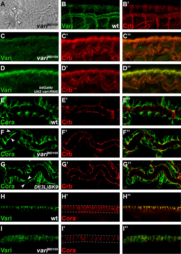
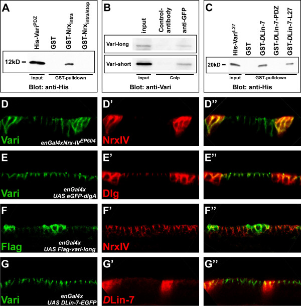
Results: Here we show by electron microscopic analysis that Drosophila embryos lacking varicose function fail to develop septate junctions in the tracheae and the epidermis. In the embryo and in imaginal discs varicose expresses two protein isoforms, which belong to the MAGUK family. The two isoforms can be distinguished by the presence or absence of two L27 domains and are differentially affected in different varicose alleles. While the short isoform is essential for viability, the long isoform seems to have a supportive function. Varicose proteins co-localise with Neurexin IV in pleated septate junctions and are necessary, but not sufficient for its recruitment. The two proteins interact in vitro by the PDZ domain of Varicose and the four C-terminal amino acids of Neurexin IV. Postembryonic reduction of varicose function by expressing double-stranded RNA affects pattern formation and morphogenesis of the wing and the development of normal-shaped and -sized eyes.

Conclusion: Expression of two Varicose isoforms in embryonic epithelia and imaginal discs suggests that the composition of Varicose-mediated protein scaffolds at septate junctions is dynamic, which may have important implications for the modulation of their function.


Figures

Figure 1
Figure 1
varicose encodes a MAGUK protein. (A) Exon-intron structure of the vari locus (scale bar according to FlyBase). Two different transcripts could be isolated: the vari-long transcript contains exons 1, 2, 3 to 10 with the translational start residing in exon 1; vari-short consists of exons 3a, 3 to 10 with the translational start site in exon 4 (dark grey boxes: exons common to both transcripts, light grey boxes: exons specific to vari-long, white boxes: UTR). Mobilisation of the P(XP)-element d10880 yielded the vari-short-specific mutant allele variMD109, which removes exon 3a and adjacent 3'-intronic DNA (shaded box). Primer pairs vari-long-5/3 and vari-short-5/3 were used to detect the corresponding vari transcripts in the wild-type (D) and different vari alleles (see Fig. 5). (B) Structure and size comparison of the MAGUK proteins Vari-long and Vari-short with their human homologs hsMPP6_c (GenBank accession number EAW93810), hsMPP6_a (GenBank accession number EAW93808) and hsMPP2_b (GenBank accession number EAW51656), respectively. The percentages of amino acid identities of the domains with respect to the corresponding domains of Vari-long are shown. (C) Northern blot of 5 μg poly(A+) RNA from staged wild-type embryos (1 = 0–4 h, 2 = 4–12 h, 3 = 12–24 h) hybridised with a probe that detects both vari-long and vari-short transcripts. (D) RT-PCR on total RNA from wild-type embryos (> 8 h old) with primer pairs vari-long-5/3 and vari-short-5/3 detects single vari-long- and vari-short-transcripts. (E) Western blot analysis of protein lysates from staged wild-type embryos and embryos of different genotypes (> 8 h old), probed with an anti-Vari antibody that recognises both Vari-long and Vari-short (1 = wt, 0–4 h, 2 = wt, 4–12 h, 3 = wt, 12–24 h, 4 = daG32>UAS Flag-vari-long, 5 = daG32>UAS vari-RNAi, 6 = Df(2L)DS6). Overexpressed Flag-Vari-long in lane 4 is slightly bigger than endogenous Vari-long due to the N-terminal Flag-tag. The protein amount loaded per lane equals 5 embryos with the exception of lysates from daG32>UAS Flag-vari-long embryos (equals 0.5 embryos). The lysate from Df(2L)DS6 embryos serves as a negative control and an unspecific, cross-reacting band (asterisk) as a loading control.
Figure 2
Figure 2
Varicose localises to septate junctions in the embryo. (A-B) A wild-type embryo of stage 15, in different focal planes, stained with anti-Vari (green) and anti-Fasciclin III (red). Vari is expressed in many epithelia of ectodermal origin. sg: salivary glands, tr: tracheae, ep: epidermis, hg: hindgut. (C-E) Epidermis of stage 15 wild-type embryos, stained with anti-Vari (green, C-E), anti-Crb (C'), anti-Arm (D'), and anti-NrxIV (E') (red), respectively. C"-E" show the merged images. Vari co-localises with the septate junction component NrxIV, basal to Crb and Arm. In A-B anterior is left and dorsal is up. In C-E apical is up.
Figure 3
Figure 3
Varicose is required for correct tracheal tube and epidermis formation. (A) Cuticle preparations of variMD109 mutant embryos exhibit convoluted tracheae (white arrows). (B, B') Wild-type embryo of stage 15 stained with anti-Vari (green) and anti-Crb (red). Vari localises at the SJ, basal of Crb. Wild-type tracheae appear straight in contrast to the convoluted tracheae in A. (C) variMD109 mutant embryo of stage 15 stained with anti-Vari (green) and anti-Crb (red). Vari is lost from the tracheae and the epidermis, while apical Crb is not affected. Tracheae appear convoluted. (D) Stage 15 embryo with targeted knockdown of vari in the tracheae of embryos by using btlGal4 (btlGal4>UAS vari-RNAi), stained with anti-Vari (green) and anti-Crb (red). Vari is reduced to background levels in the tracheae, but not affected in the epidermis. Apical localisation of Crb is not affected in the tracheae. (E) Dorsal tracheal trunk of a wild-type embryo of stage 15, stained with anti-Coracle (Cora; green) and anti-Crb (red). Cora localises in the SJ, basal to Crb. (F) Dorsal tracheal trunk of a variMD109 mutant embryo of stage 15, stained with anti-Cora (green) and Crb (red). Cora is delocalised to apical and basal sites (white arrows), whereas Crb remains in its apical position. (G) Dorsal tracheal trunk of a Df(3L)BK9 mutant embryo of stage 15, in which the NrxIV locus is deleted, stained with anti-Cora (green) and anti-Crb (red). As in variMD109 mutant embryos, Cora becomes mislocalised to apical and basal positions (white arrows) in the absence of NrxIV, while apical localisation of Crb is not affected. (H) Epidermis of a wild-type embryo of stage 15, stained with anti-Vari (green) and anti-Cora (red). Both proteins are co-localised at the SJ. (I) variMD109 mutant embryo of stage 15, stained with anti-Vari (green) and anti-Cora (red). The amount of Cora is reduced and the remaining Cora protein is mislocalised along the whole lateral membrane. In B-D and H-I apical is up. White dotted lines in H' and I' mark the apical and basal side of the epithelial cells, respectively.
Figure 4
Figure 4
EM analysis reveals defective septate junctions in vari mutants. Epidermis (A-D, I) and tracheae (E-H) of stage 16 embryos. (A, E): variMD109/CyO-twi-GFP; (B, F): variMD109; (C, G): vari03953b/CyO-twi-GFP; (D, H): vari03953b; (I): wild-type. Adherens junctions (black arrowheads) and developing septate junctions (white arrows) can be distinguished in wild-type embryos. The inset in E shows the regularly aligned septa of pleated septate junctions between adjacent cells. Septate junctions are absent in homozygous variMD109 (B, F) and vari03953b (D, H) mutant embryos, while adherens junctions are well developed (black arrowheads).
Figure 5
Figure 5
RT-PCR- and western blot-analysis of vari-mutant alleles. (A) RT-PCR on total RNA of wild-type and different vari-mutant embryos (> 8h), using vari-long- and vari-short-specific primer pairs vari-long-5/3 or vari-short-5/3 (see Fig. 1A). No vari-long transcripts can be detected in embryos homozygous for the deficiency Df(2L)DS6 and the allele variF033. vari-long transcripts are slightly more abundant in variMD109 and strongly enriched in vari03953b embryos. Both vari-long and vari-short transcripts are truncated in vari38EFa2 (black arrowheads). vari-short transcripts are absent in variMD109, strongly reduced in variF033 and vari03953b, but only mildly affected in variR3, vari327 and variR979. Transcripts from the rp49 gene serve as internal loading control. (B) Western blot analysis of the same mutant alleles as in A. Vari-long protein is present in wild-type amounts in variMD109, absent in embryos homozygous for the deficiency Df(2L)DS6 and the allele variF033, reduced in vari03953b,vari327 and variR979 and truncated in variR3 (white arrow) and vari38EFa2 (asterisk). Vari-short protein is absent in variMD109, in the deficiency Df(2L)DS6 and in variR3 and reduced in the remaining alleles. RT-PCR and western-blot analysis of the deficiency Df(2L)DS6, which removes the vari locus, serves as negative control.
Figure 6
Figure 6
Vari interacts with NrxIV in vitro and in vivo. (A) A GST-NrxIV fusion protein comprising the entire intracellular domain of NrxIV (including the PDZ-binding motif-EIFI) pulls down a His-tagged Vari transgene that contains only the PDZ domain (Pro169-Val270) in vitro. However, a GST-NrxIV fusion protein including a C-terminally truncated intracellular domain of NrxIV (lacking the PDZ-binding motif-EIFI) does not. (B) Co-immunoprecipitation assay from embryonic protein extracts (> 8 h; genotype: NrxIV-GFP/TM6B) reveals that in the embryo NrxIV forms a complex with Vari-long and Vari-short. (C) Both GST-DLin-7 and GST-DLin-7-L27 fusion protein (containing only the L27 domain of DLin-7), pull down the His-tagged Vari N-terminus (Ser5-Tyr157), which comprises the putative L27 domains, while GST-DLin-7-PDZ fails to pull down Vari. (D-G) Wild-type embryos of stage 15 overexpressing different UAS-transgenes in a striped pattern using enGal4. Green: anti-Vari, Red: anti-NrxIV (D, F); Green: anti-Vari, Red: anti-Dlg (E); Green: anti-Vari, Red: anti-DLin-7 (G). Recruitment of Vari by ectopic NrxIV is specific. In D-G apical is up.
Figure 7
Figure 7
RNAi-mediated knockdown of vari in the wing imaginal disc. (A) Gal4/UAS-mediated overexpression of a vari-RNAi transgene along the antero-posterior boundary of a third instar wing imaginal disc with ptcGal4 significantly reduces the amount of Vari protein (white arrows). (B) Close-up view on imaginal disc cells from the wing imaginal disc shown in A. In the overexpression region (dotted white line in B) Vari (green) is no longer detectable at the SJ. Crb (red) still localises to the SAR, demonstrating that apico-basal polarity of the wing imaginal disc epithelial cells is unaffected. (C) Wing blades from adult wild-type flies (C) and flies in which the vari-RNAi transgene was expressed with sdGal4; Gbe+Su(H)lacZ. Overexpression of the transgene affects wing shape and margin formation to different degrees. Bristles and hairs are lost from the posterior and, more rarely, anterior wing margin, respectively (arrows in C', C"). The wings of adult sdGal4; Gbe+Su(H)lacZ>UAS vari-RNAi flies normally appear 'collapsed', but unfold again during the embedding procedure. (D) Anti-Wg antibody staining of sdGal4; Gbe+Su(H)lacZ (D) and sdGal4; Gbe+Su(H)lacZ>UAS vari-RNAi (D', D") third instar wing imaginal discs. In comparison to wild-type (D) vari knockdown leads to a lighter and discontinuous Wg expression at the prospective wing margin (arrows in D', D"). (E) X-Gal staining of sdGal4; Gbe+Su(H)lacZ (E) and sdGal4; Gbe+Su(H)lacZ>UAS vari-RNAi (E') third instar wing imaginal discs to visualize activity of the Notch signaling pathway. Notch activation at the prospective wing margin appears to be more diffuse and sometimes interrupted (arrow in E'). (F) Western blot analysis of protein lysates from wild-type wing and eye imaginal discs, wild-type ovaries as well as wild-type and Df(2L)DS6 embryos, probed with an anti-Vari antibody that recognises both Vari-long and Vari-short. Whereas in wing and eye imaginal discs both Vari-long and Vari-short can be detected, wild-type ovaries express only Vari-short. The protein amount loaded per lane equals 10 wing or eye imaginal discs, 1 ovary and 5 embryos, respectively. The lysates from wild-type and Df(2L)DS6 embryos serve as controls and the unspecific, cross-reacting band slightly below 72K as a loading control. In A, B, D and E anterior is left and dorsal is up. In C anterior is up.
Figure 8
Figure 8
Vari function in the developing eye. (A, A') Third instar wild-type eye imaginal discs stained with anti-Vari (green). Vari is predominantly expressed behind the morphogenetic furrow (white arrow in A) in the developing ommatidia. The close-up view on ommatidia of the 4-cone cell stage (A') demonstrates that Vari localises to the lateral membranes of the photoreceptor and cone cells. (B, B') Close-up views of ommatidia, stained with antibodies against Cora (B, green) and NrxIV (B', red), respectively. Cora and NrxIV are expressed in the photoreceptor and cone cells. (C-C') Head of a wild-type fly (C) and a fly overexpressing vari-RNAi with eyGal4 (C'). Reduction of vari induces roughening, downsizing and malformation of the eye. (D-D') Eye imaginal discs of third instar wild-type larvae (D) and larvae overexpressing vari-RNAi with eyGal4 (D'), stained with anti-Futsch/22C10 antibody to mark the photoreceptor cells. Reduction of vari function (D') leads to aberrant folding of the eye imaginal disc and additional tissue formation at the antennal disc (arrow). Black arrowheads mark the position of the morphogenetic furrow in D. bw = Bolwig's nerve, ad = antennal disc, ed = eye disc. (E-E") X-Gal staining of eyGal4; Gbe+Su(H)lacZ (E) and eyGal4; Gbe+Su(H)lacZ>UAS vari-RNAi (E') third instar eye imaginal discs to visualize activity of the Notch signaling pathway. Despite morphological aberrations Notch activity does not seem to be strikingly affected by RNAi-mediated knockdown of vari. eyGal4; Gbe+Su(H)lacZ>UAS vari-RNAi eye imaginal discs from second instar larvae already display abnormal folding (E", arrow). The eye imaginal disc in E" is shown in a higher magnification than those in E and E'. (F, F') Semi-thin sections of eyes from wild-type flies (F) and flies overexpressing vari-RNAi with eyGal4 (F'). In ommatidia with reduced vari function (F') the seven photoreceptor cells visible do not exhibit major morphogenetic defects and are still organised in a trapezoid pattern as in wild-type (F). In A, D and E anterior is left.

References

    1. Funke L, Dakoji S, Bredt DS. Membrane-associated guanylate kinases regulate adhesion and plasticity at cell junctions. Annu Rev Biochem. 2005;74:219–245. doi: 10.1146/annurev.biochem.74.082803.133339. - DOI - PubMed
    1. te Velthuis AJW, Admiraal JF, Bagowski CP. Molecular evolution of the MAGUK family in metazoan genomes. BMC Evol Biol. 2007;7:129–139. doi: 10.1186/1471-2148-7-129. - DOI - PMC - PubMed
    1. Conte I, Lestingi M, den Hollander AI, Miano MG, Alfano G, Circolo D, Pugliese M, Testa F, Simonelli F, Rinaldi E, Baiget M, Banfi S, Ciccodicola A. Characterisation of MPP4, a gene highly expressed in photoreceptor cells, and mutation analysis in retinitis pigmentosa. Gene. 2002;297:33–38. doi: 10.1016/S0378-1119(02)00872-7. - DOI - PubMed
    1. Mendoza C, Olguín P, Lafferte G, Thomas U, Ebitsch S, Gundelfinger ED, Kukuljan M, Sierralta J. Novel isoforms of Dlg are fundamental for neuronal development in Drosophila. J Neurosci. 2003;23:2093–2101. - PMC - PubMed
    1. Bachmann A, Timmer M, Sierralta J, Pietrini G, Gundelfinger ED, Knust E, Thomas U. Cell type-specific recruitment of Drosophila Lin-7 to distinct MAGUK-based protein complexes defines novel roles for Sdt and Dlg-S97. J Cell Sci. 2004;117:1899–1909. doi: 10.1242/jcs.01029. - DOI - PubMed

Publication types

MeSH terms

LinkOut - more resources