Comparative Study
. 2008 Jul 20;336(1):71-7.
doi: 10.1016/j.jim.2008.04.003. Epub 2008 Apr 28.

Screening monoclonal antibodies for cross-reactivity in the ferret model of influenza infection

Affiliations
Comparative Study

Screening monoclonal antibodies for cross-reactivity in the ferret model of influenza infection

John A Rutigliano et al. J Immunol Methods. .

Abstract

Influenza virus infections carry a high public health cost, and pandemics are potentially catastrophic. Though the ferret is generally regarded as the best model for human influenza, few reagents are available for the analysis of cellular immunity. We thus screened monoclonal antibodies (mAbs) made for identifying immune cells in other species to see if any were cross-reactive. Flow cytometric analysis of lymphocytes isolated from blood, spleen, and lung of normal and virus-infected ferrets indicated that several mouse mAbs bound to the corresponding antigens in ferrets. Typing bronchoalveolar lavage populations from pneumonic ferrets with mAb to human CD8 showed the massive CD8+ T cell enrichment characteristic of this infection in mice. The availability of this, and several other mAbs that showed cross-reactivity, should allow us to begin the dissection of cell-mediated immunity in the ferret, which, at least from these early results, looks similar to the situation in mice.


Figures

Figure 1
Figure 1
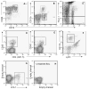
Staining profiles for mAbs that showed evidence of cross-reactivity (see Table 1). The PBMCs were from ferrets that had been previously exposed to influenza A virus. A) Histogram and dot plot for staining with the anti-human CD8 clone OKT8. B) Histogram and dot plot staining with anti-mouse CD11b. C-G) Dot plots indicating cross-reactivity with anti-mouse CD44 (C), Thy1.1 (D), CD43 (E), LFA-1 (F), and CD103 (G)
Figure 2
Figure 2
Surface antigen staining for lymphocytes recovered by BAL of influenza A virus-infected ferrets. The ferrets were first primed with either an H1N1 or H2N3 influenza virus. Several weeks later, they were challenged i.n. with Wuhan H3N2, seven days prior to sampling. A-C) Dot plots of OKT8 (A-C), CD44 (A), CD11b (B), and Thy1.1 (C), which were also cross-reactive on PBMCs in Fig.1. Additional cross-reactivities seen with anti-mouse CD25 (D), CD43 (E), Ly6C (F), and LFA-1 (G) are shown in the subsequent panels. A dot plot representing unstained cells from the ferret BAL is shown in panel H. Results from staining of splenocytes are not shown but are similar to what was seen in cells from the BAL.
Figure 3
Figure 3
Frequency values for surface antigen positive cells recovered from lung or spleen of influenza virus A-infected ferrets. The percentage of cells staining in BAL (A) and spleen (B) populations is shown for the experiment described in the legend to Figure 2. One ferret that was primed with H1N1 went unchallenged. Spleen cells from a previously-infected mouse were used as previously described (Keating et al., 2007) as a positive control for the mouse mAbs (B).
Figure 4
Figure 4
Intracellular staining profiles for PMA + ionomycin-stimulated cells from the experiment described in Figure 2. A) Dot plots for staining of cells from ferret BAL with mAbs to bovine IFN-γ and IL-4 that have been previously shown to cross-react with mink. B) Profiles for IFN-γ and TNF in the spleen using anti-mouse antibodies. C-D) Graphs showing the percentage of OKT8+ T cells that were positive for the indicated cytokine in the BAL (C) and spleens (D). Cells from a single mouse spleen were used as a positive control for the mouse mAbs (D).

References

    1. Aasted B. Mink infected with Aleutian Disease Virus have an elevated level of CD8-positive T lymphocytes. Vet Immunol Immunopath. 1989;20:375–385. - PubMed
    1. Brodersen R, Bijlsma F, Gori K, Jensen KT, Chen W, Dominguez J, Haverson K, Moore PF, Saalmuller A, Sachs D, Slierendrecht WJ, Stokes C, Vainio O, Zuckerman F, Aasted B. Analysis of the immunological cross reactivities of 213 well characterized monoclonal antibodies with specificities against various leucocyte surface antigens of human and 11 animal species. Vet Immunol Immunopath. 1998;64:1–13. - PubMed
    1. Cabezas JA, Calvo P, Eid P, Martin J, Perez E, Reglero A, Hannoun C. Neuraminidase from influenza A virus A (H3N2): specificity towards several substrates and procedure of activity determination. Biochim Biphys Acta. 1980;616:228–238. - PubMed
    1. Campbell DG, Gagnon J, Reid K, Williams AF. Rat brain Thy-1 glycoprotein. The amino acid sequence, disulphide bonds and an unusual hydrophobic region. Biochem J. 1981;195:15–30. - PMC - PubMed
    1. Chan MCW, Cheung CY, Chui WH, Tsao SW, Nicholls JM, Chan YO, Chan R, Long HT, Poon L, Guan Y, Peiris JSM. Proinflammatory cytokine responses induced by influenza A (H5N1) viruses in primary human alveolar and bronchial epithelial cells. Resp Res. 2005;6:135–147. - PMC - PubMed

Publication types

MeSH terms

Substances