Review
. 2008 Jun 25;288(1-2):95-103.
doi: 10.1016/j.mce.2008.04.012. Epub 2008 Apr 26.

Gdnf signaling pathways within the mammalian spermatogonial stem cell niche

Affiliations
Review

Gdnf signaling pathways within the mammalian spermatogonial stem cell niche

Marie-Claude Hofmann. Mol Cell Endocrinol. .

Abstract

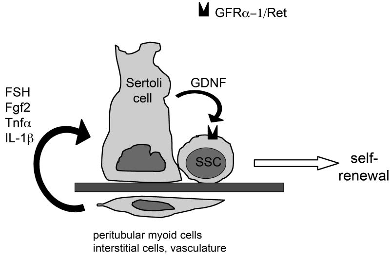
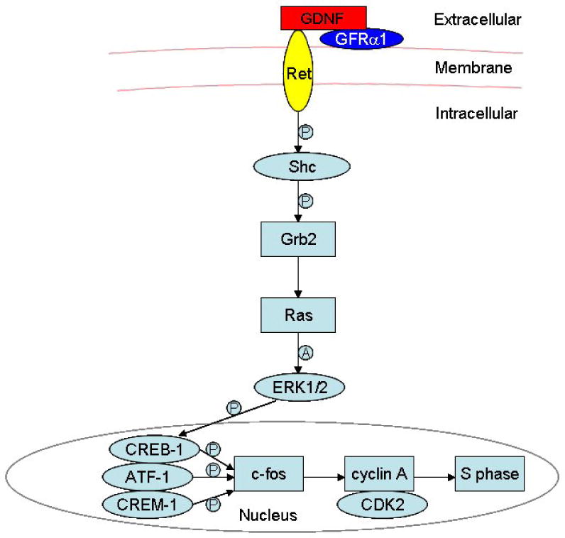
Mammalian spermatogenesis is a complex process in which male germ-line stem cells develop to ultimately form spermatozoa. Spermatogonial stem cells, or SSCs, are found in the basal compartment of the seminiferous epithelium. They self-renew to maintain the pool of stem cells throughout life, or they differentiate to generate a large number of germ cells. A balance between SSC self-renewal and differentiation in the adult testis is therefore essential to maintain normal spermatogenesis and fertility. Maintenance and self-renewal are tightly regulated by extrinsic signals from the surrounding microenvironment, called the spermatogonial stem cell niche. By physically supporting the SSCs and providing them with growth factors, the Sertoli cell is the main component of the niche. In addition, adhesion molecules that connect the SSCs to the basement membrane and cellular components of the interstitium between the seminiferous tubules are important regulators of the niche function. This review mainly focuses on glial cell line-derived neurotrophic factor (Gdnf), which is produced by Sertoli cells to maintain SSCs self-renewal, and the downstream signaling pathways induced by this crucial growth factor. Interactions between Gdnf and other signaling pathways that maintain self-renewal, as well as the role of novel SSC- and Sertoli cell-specific transcription factors, are also discussed.


Figures

Figure 1
Figure 1. The first steps of mammalian spermatogenesis
Diagram representing the first steps of spermatogenesis, in particular the different subtypes of A spermatogonia. The spermatogonial stem cell (SSC or As or Asingle spermatogonium is able to self-renew (curved arrow) or to differentiate into Apaired spermatogonia linked by an intercellular bridge (straight arrow). The Apaired spermatogonia subsequently proliferate into 4, 8, and 16 Aaligned spermatogonia, ultimately producing differentiating type A spermatogonia. The differentiating type A spermatogonia proliferate and differentiate to become spermatocytes that undergo meiosis, producing spermatids that will go through spermiogenesis. All spermatogenic cells differentiate as cohorts of units interconnected by intercellular bridges. Adapted from Kiger and Fuller, Male Germline Stem Cells. In: Stem Cell Biology, Cold Spring Harbor Laboratory Press, 2001 (Kiger and Fuller, 2001).
Figure 2
Figure 2. Organization of the Drosophila testis and the seminiferous epithelium in mammals
Figure 2A: Germinal proliferation center in Drosophila. In the apical tip region, the germline stem cells (S) are in contact with the hub cells (H). Each germline stem cell also associates with a pair of somatic cells called cyst progenitor cells (CP). When a germline stem cell or a cyst progenitor cell divides, the daughter cell that loses contact from the hub differentiates into a gonialblast (G) or a cyst cell (C) respectively. Two cyst cells will associate with one gonialblast and will never divide again. They will enclose the progeny of the gonialblast throughout spermatogenesis. From Fuller MT, Seminars in Developmental Biology, 1998 (Fuller, 1998). Figure 2B: Seminiferous epithelium of the mammalian testis. The spermatogonial stem cells are a subpopulation of type A spermatogonia that reside in the basal compartment of the seminiferous epithelium, in contact with the basement membrane. As the germ cells proliferate and differentiate, they move toward the lumen of the seminiferous tubule. Adapted from Russell L, Mammalian Spermatogenesis, In: Histological and Histopathological Evaluation of the Testis, Cache River Press, 1990 (Russell, 1990).
Figure 3
Figure 3. Regulation of GDNF expression in Sertoli cells
In the mouse testis, the expression of GDNF by Sertoli cells is under control of FSH, FGF2, Tnfα and IL-1β, probably produced by interstitial cells. Therefore, SSCs self-renewal is controlled both systemically and locally.
Figure 4
Figure 4. GDNF/Src signaling in spermatogonial stem cells
This model shows how GDNF can promote cell cycle progression via activation of Src kinase(s) and a PI3K/Akt pathway to increase N-myc gene expression. All 4 kinases depicted are inhibited by SU6656 and are involved in spermatogonial proliferation. p60-Src and c-Yes are necessary for the early response to GDNF, while the addition of Lyn and Fyn might be important for proliferation associated with differentiation. From Braydich-Stolle et al, Dev Biol, 2007 (Braydich-Stolle et al., 2007).
Figure 5
Figure 5. Immunocytochemistry study showing the up-regulation of nuclear N-Myc expression after stimulation by GDNF
a: C18-4 spermatogonial stem cell line with 10% Nu serum in the culture media, negative control (no first antibody); b: C18-4 cells with 10% Nu serum in the culture media, showing a weak, basal expression of N-Myc; c: C18-4 cells with 10% Nu serum and 100 ng/ml GDNF in the culture media, showing an increase in staining intensity for N-Myc in comparison to the basal expression; d: cluster of primary spermatogonial stem cells grown for 3 days in presence of GDNF (100 ng/ml) and showing strong up-regulation of N-myc only in some cells (arrows). Because GDNF activates N-Myc expression in a subpopulation of undifferentiated spermatogonia, it suggests that N-Myc activation is relevant for spermatogonial stem cell self-renewal only. From Braydich-Stolle et al, Dev Biol, 2007 (Braydich-Stolle et al., 2007).
Figure 6
Figure 6. GDNF/Ras signaling in spermatogonial stem cells
The schematic diagram demonstrates intracellular signaling events in the Ras/ERK1/2 pathway as well as the downstream cascades activated by GDNF in spermatogonial stem cells. “P” indicates “phosphorylate”, and “A” denotes “activate”. From He et al, Stem Cells, 2008 (He et al., 2008).
Figure 7
Figure 7. Regulation of Etv5 expression in Sertoli cells
In the mouse testis, the expression of the transcription factor Etv5 in Sertoli cells is under the control of Fgf2 and Egf, probably produced by interstitial cells. Genes regulated by Etv5 might include chemokines important for SSC homing.

References

    1. Abdallah B, Hourdry J, Krieg P, Denis H, Mazabraud A. Germ cell-specific expression of a gene encoding eukaryotic translation elongation factor 1 alpha (eEF-1 alpha) and generation of eEF-1 alpha retropseudogenes in Xenopus laevis. Proc Natl Acad Sci U S A. 1991;88:9277–9281. - PMC - PubMed
    1. Airaksinen M, Saarma M. The GDNF family: signalling, biological functions and therapeutic value. Nat Rev Neurosci. 2002;3:383–394. - PubMed
    1. Barna M, Hawe N, Niswander L, Pandolfi P. Plzf regulates limb and axial skeletal patterning. Nat Genet. 2000;25:166–172. - PubMed
    1. Braydich-Stolle L, Kostereva N, Dym M, Hofmann M. Role of Src family kinases and N-Myc in spermatogonial stem cell proliferation. Dev Biol. 2007;304:34–45. - PMC - PubMed
    1. Braydich-Stolle L, Nolan C, Dym M, Hofmann M. Role of glial cell line-derived neurotrophic factor in germ-line stem cell fate. Ann N Y Acad Sci. 2005;1061:94–99. - PMC - PubMed

Publication types

Substances