

Changes mimicking new leptomeningeal disease after intensity-modulated radiotherapy for medulloblastoma
- PMID: 18485616
- PMCID: PMC2953789
- DOI: 10.1016/j.ijrobp.2008.03.056
Changes mimicking new leptomeningeal disease after intensity-modulated radiotherapy for medulloblastoma
Abstract
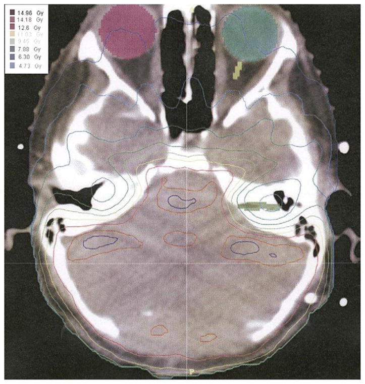
Purpose: Acute and late changes in magnetic resonance imaging of the pediatric brain have been described after radiotherapy (RT). We report the post-RT neuroimaging changes in the posterior fossa after intensity-modulated RT (IMRT) in children with medulloblastoma and contrast them with those of leptomeningeal disease.
Methods and materials: We performed a retrospective review of 53 consecutive children with medulloblastoma who were treated with craniospinal RT followed by IMRT to the posterior fossa and chemotherapy between 1997 and 2006.
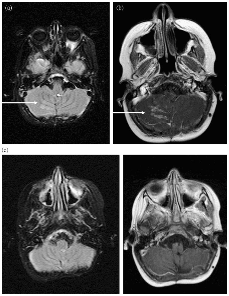
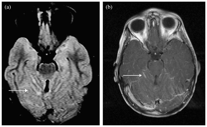
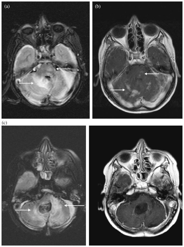
Results: After IMRT to the posterior fossa, 8 (15%) of 53 patients developed increased fluid-attenuated inversion-recovery signal changes in the brainstem or cerebellum and patchy, multifocal, nodular contrast enhancement at a median of 6 months. The enhancement superficially resembled leptomeningeal disease. However, the enhancement resolved without intervention at a median of 6 months later. The accompanying fluid-attenuated inversion-recovery signal changes occasionally preceded the enhancement, were often parenchymal in location, and resolved or persisted to a lesser degree. All 8 patients with transient magnetic resonance imaging changes in the posterior fossa were alive at last follow-up. In contrast, leptomeningeal disease occurred in 8 (15%) of our 53 patients at a median of 19.5 months after IMRT completion. Of these 8 patients, 7 demonstrated initial nodular enhancement outside the conformal field, and 7 patients died.
Conclusion: Magnetic resonance imaging changes can occur in the posterior fossa of children treated with IMRT for medulloblastoma. In our experience, these transient changes occur at a characteristic time and location after RT, allowing them to be distinguished from leptomeningeal disease.
Conflict of interest statement
Conflict of interest: none.
Figures
References
-
- Blaney SM, Kun LE, Hunter J, et al. Tumors of the central nervous system. In: Pizzo PA, Poplack DG, editors. Principles and practice of pediatric oncology. 5. Philadelphia, PA: Lippincott Williams & Wilkins; 2006. pp. 818–825.
-
- Teh BS, Woo SY, Butler EB. Intensity modulated radiation therapy (IMRT): A new promising technology in radiation oncology. Oncologist. 1999;4:433–442. - PubMed
-
- Hertzberg H, Huk WJ, Ueberall MA, et al. for the German Late Effects Working Group. CNS late effects after ALL therapy in childhood: Part I. Neurological findings in long-term survivors of childhood ALL—An evaluation of the interferences between morphology and neuropsychological performance. Med Pediatr Oncol. 1997;28:387–400. - PubMed
-
- Bakardjiev AI, Barnes PD, Goumnerova LC, et al. Magnetic resonance imaging changes after stereotactic radiation therapy for childhood low grade astrocytoma. Cancer. 1996;78:864–873. - PubMed
-
- Helton KJ, Edwards M, Steen G, et al. Neuroimaging-detected late transient treatment-induced lesions in pediatric patients with brain tumors. J Neurosurg. 2005;102(Suppl 2):179–186. - PubMed
Publication types
MeSH terms
Grants and funding
LinkOut - more resources
Full Text Sources
Medical
