. 2008 May 16;133(4):612-26.
doi: 10.1016/j.cell.2008.03.025.

Acetylation is indispensable for p53 activation

Affiliations

Acetylation is indispensable for p53 activation

Yi Tang et al. Cell. .

Erratum in

  • Cell. 2008 Jun 27;133(7):1290

Abstract

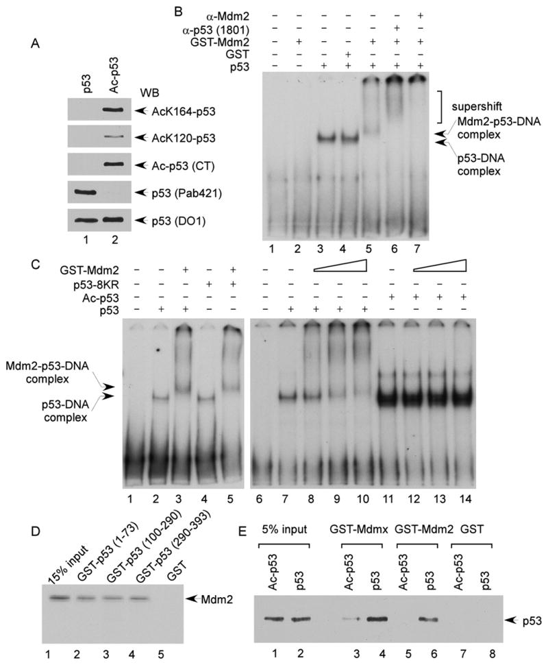
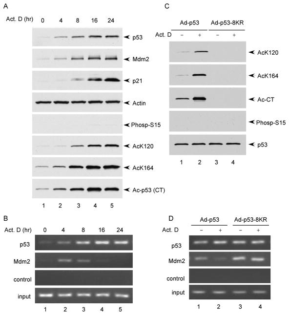
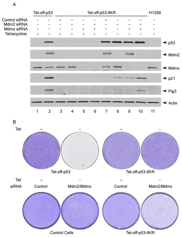
The activation of the tumor suppressor p53 facilitates the cellular response to genotoxic stress; however, the p53 response can only be executed if its interaction with its inhibitor Mdm2 is abolished. There have been conflicting reports on the question of whether p53 posttranslational modifications, such as phosphorylation or acetylation, are essential or only play a subtle, fine-tuning role in the p53 response. Thus, it remains unclear whether p53 modification is absolutely required for its activation. We have now identified all major acetylation sites of p53. Although unacetylated p53 retains its ability to induce the p53-Mdm2 feedback loop, loss of acetylation completely abolishes p53-dependent growth arrest and apoptosis. Notably, acetylation of p53 abrogates Mdm2-mediated repression by blocking the recruitment of Mdm2 to p53-responsive promoters, which leads to p53 activation independent of its phosphorylation status. Our study identifies p53 acetylation as an indispensable event that destabilizes the p53-Mdm2 interaction and enables the p53-mediated stress response.


Figures

Figure 1
Figure 1. Identification of K164 within the Human p53 DNA-Binding Domain as a Novel Acetylation Site by p300/CBP
(A) Schematic representation of the human p53 protein with known acetylation sites indicated, including K120, K370, K372, K373, K381, K382, and K386. In this study, K164 was identified to be acetylated by p300/CBP. (B) Mass spectrometry analysis of the p53-derived peptides containing acetylated K164 (AcK164). The protein was prepared as described in the Experimental Procedures. (C) Alignment of the K164 flanking region of the human p53 protein with those of p53 from other species and of human p63 and p73. The conserved lysine residue is marked in bold; h: human; m: mouse; c: chicken; x: Xenopus; and z: zebrafish. (D) In vitro acetylation of p53 by p300/CBP. The Flag-p53-K120R/6KR (lanes 2, 4, and 6) or Flag-p53-K120R/K164/6KR (lanes 1, 3, and 5) recombinant protein was incubated alone (lanes 1 and 2), with p300 (lanes 3 and 4), or with CBP (lanes 5 and 6). The reaction products were resolved by SDS-PAGE and analyzed by western blot using the site-specific polyclonal antibody against acetylated K164 (anti-AcK164-p53) (top panel). The levels of the p53 recombinant protein substrates are shown in the bottom panel by Ponceau red staining. (E) Acetylation of the endogenous p53 protein at K164. HCT116 cells were treated with 20 μM etoposide (0, 4, and 6 hr). The endogenous p53 proteins were immunoprecipitated by the anti-p53 (1801) antibody and assayed for acetylation by western blot using the anti-AcK164-p53 antibody (top panel). The levels of p53 in the cell extracts are shown in the middle panel.
Figure 2
Figure 2. Functional Characterization of Acetylation-Defective p53 Mutants
(A) Scheme of the human p53 lysine-to-arginine mutants used in this study. (B) Induction of p21 and Mdm2 by p53 lysine-to-arginine mutants. H1299 cells were transfected with increasing amount of plasmid DNA expressing different p53 mutants. The total cell extracts were subjected to western blot for p53, p21, and Mdm2, and GFP was used as a transfection control. (C) As in (B), two additional p53 lysine-to-arginine mutants were examined. (D) In vitro DNA-binding activities of p53 and p53-8KR. Gel shift assay was performed using a fragment containing a p53-binding site from the p21 promoter, as described in the Experimental Procedures. (E) Degradation of the p53 and p53-8KR proteins by Mdm2. H1299 cells were cotransfected with CMV-p53 expressing either p53 or p53-8KR and an increased amount of CMV-Mdm2. The cellular levels of p53 were determined by western blot using anti-p53 antibody (DO-1).
Figure 3
Figure 3. Lack of Acetylation at Its DNA-Binding Domain and C Terminus Abolishes p53 Ability to Induce Cell Growth Arrest
(A) Expression of the human p53 protein at physiological levels in Tet-off-p53 H1299 cells. The total cell extracts from Tet-off-p53 cells (before and after induction) and HCT116 cells (treated without and with etoposide) were assayed by western blot using the antibodies against p53 (DO-1) and actin. (B) Acetylation of p53 in Tet-off-p53 cells. The Flag-p53 proteins enriched from Tet-off-p53 and Tet-off-p53-8KR cells by M2 immunoprecipitation were subjected to western blot analysis using acetylation- and phosphorylation-specific antibodies, as indicated, and anti-p53 (1801) to determine the levels of total p53. (C) Tet-off-p53 and Tet-off-p53-8KR cells were induced for 0, 1, 2, 3, and 4 days. The total cell extracts were analyzed by western blot using antibodies against p53 (DO-1), Mdm2, Bax, Puma, Pig3, p21, and actin. (D) Four days after induction, Tet-off-p53 and Tet-off-p53-8KR cells were treated with 10 μM Brdu for 1 hr and immunostained with the anti-BrdU antibody. The nuclei are in blue (DAPI), and Brdu-positive nuclei are shown in red. (E) Tet-off-p53 and Tet-off-p53-8KR cells were induced as in (C), and incorporation of BrdU was assayed as in (D).
Figure 4
Figure 4. Acetylation of p53 Abrogates the Promoter-Specific Recruitment of Mdm2 and Mdmx
(A) H1299 cells transfected by various plasmids, as indicated, were treated with 1% formaldehyde for 10 min and processed for ChIP analysis. The occupancy of p53, Mdm2, CBP, and Tip60 of the p21 promoter was detected by PCR. (B) Acetylation of p53 in the presence of CBP and Tip60. H1299 cells were transfected as in (A), and Flag-p53 immunoprecipitated with M2 beads was assayed for acetylation at specific sites by western blot. Expression of Mdm2, CBP, and Tip60 was detected in the total cell extracts. (C) H1299 cells transfected by the indicated plasmids were treated with 1% formaldehyde for 10 min and processed for ChIP analysis as in (A). The occupancy of p53, Mdm2, Mdmx, CBP, and Tip60 of the p21, Pig3, and Mdm2 promoters was detected by PCR-agarose gel electrophoresis. The relative levels of recruitment of Mdm2 and Mdmx were further quantitated by real-time PCR as shown in Figures S7, S8, and S9.
Figure 5
Figure 5. Acetylation Disrupts the p53-Mediated Mdm2-p53-DNA Complex Formed In Vitro
(A) The fully acetylated Flag-p53 proteins were purified from H1299 cells cotransfected with plasmid DNA expressing CMV-Flag-p53, CMV-Tip60, and CBP-HA. Acetylation levels were examined by western blot using site-specific acetylation antibodies, as indicated. (B) Gel shift assay was performed, according to the Experimental Procedures, to analyze Mdm2-p53-DNA complex. A 32P-labeled 159 bp DNA fragment containing the p53-binding site from the human p21 promoter was incubated with proteins, as indicated. The reaction was resolved by 4% PAGE and protein-DNA complexes were visualized by autoradiography. (C) Gel shift assays were carried out as in (B). The DNA probe was incubated with the Flag-p53, Flag-p53-8KR, or acetylated Flag-p53 proteins (20 ng) in the absence or presence of an increasing amounts of GST-Mdm2. (D) In vitro binding of Mdm2 to the various domains of p53. The in vitro translated 35S-methionine-labeled Mdm2 protein was used in the pull-down assay with GST (lanes 5), GST-p53 (1–73) (lane 2), GST-p53 (100–290) (lane 3), or GST-p53 (290–393) (lane 4). The glutathione elutes and 15% input materials (lane 1) were resolved in SDS-PAGE gel and visualized by autoradiography. Levels of the recombinant proteins were shown in the Figure S6B. (E) In the GST-pull-down assays, the purified p53 or acetylated p53 proteins were incubated with GST (lanes 7 and 8), GST-Mdm2 (lanes 5 and 6), or GST-Mdmx (lanes 3 and 4). The glutathione eluted material and 5% input materials (lanes 1 and 2) were analyzed by western blot using anti-p53 antibody (DO-1). GST fusion proteins were shown in Figures S6C and S6D.
Figure 6
Figure 6. Recruitment of Mdm2 to the p21 Promoter in Response to DNA Damage
(A) Cell extracts from U2OS cells treated with actinomycin D (10 nM) for 0, 4, 8, 16, and 24 hr were fractionated in SDS-PAGE gel and analyzed by western blot using antibodies against p53 (FL), Mdm2, ser-15-phospho-p53, and actin. Acetylation levels of p53 were determined by immunoprecipitation with site-specific acetylation antibodies followed by western blot using a monoclonal p53 antibody DO-1. (B) U2OS cells were treated as in (A) and processed for ChIP assay. The binding of p53 and Mdm2 to the p21 promoter was detected by PCR. (C) The Flag-p53 proteins were immunoprecipitated with M2 beads from the total cell extracts of H1299 cells infected with the indicated adenoviruses for 24 hr and treated with or without actinomycin D (10 nM) for 16 hr. The levels of acetylation or phosphorylation were detected by western blot using site-specific antibodies. (D) H1299 cells were infected and treated as in (C) and processed for ChIP assay. The binding of p53 and Mdm2 to the p21 promoter was detected by PCR.
Figure 7
Figure 7. Cell Growth Repression Function of p53 Is Abrogated by Loss of Acetylation but Rescued by siRNA-Mediated Ablation of Mdm2 and Mdmx
(A) Tet-off-p53 cells and Tet-off-p53-8KR cells treated with control siRNA, Mdm2 siRNA, Mdmx siRNA, or a combination of Mdm2 siRNA and Mdmx siRNA, were induced by removal of tetracycline from the media. The total cell extracts were analyzed by western blot using antibodies against p53 (DO-1), Mdmx, Mdm2, Pig3, p21, and actin. (B) In the upper panel, p53-8KR is unable to induce cell death and growth arrest. Tet-off-p53 and Tet-off-p53-8KR cells were plated at 10% confluence and incubated in the presence (uninduced) or absence (induced) of tetracycline for 6 days. The plates were washed with PBS and stained with crystal violet. In the lower panel, Tet-off-p53-8KR treated with either control siRNA or Mdm2/Mdmx siRNA were split and incubated in the presence (uninduced) or absence (induced) of tetracycline for 6 days. The plates were stained with crystal violet.

Similar articles

Cited by

References

    1. An W, Kim J, Roeder RG. Ordered cooperative functions of PRMT1, p300, and CARM1 in transcriptional activation by p53. Cell. 2004;117:735–748. - PubMed
    1. Aparicio O, Geisberg JV, Struhl K. Chromatin Immunoprecipitation for determining the association of proteins with specific genomic sequences in vivo. Curr Protoc Cell Biol. 2004;17:7.1–7.23. - PubMed
    1. Arva NC, Gopen TR, Talbott KE, Campbell LE, Chicas A, White DE, Bond GL, Levine AJ, Bargonetti J. A chromatin-associated and transcriptionally inactive p53-Mdm2 complex occurs in Mdm2 SNP309 homozygous cells. J Biol Chem. 2005;280:26776–26787. - PubMed
    1. Ashcroft M, Kubbutat MH, Vousden KH. Regulation of p53 function and stability by phosphorylation. Mol Cell Biol. 1999;19:1751–1758. - PMC - PubMed
    1. Ashcroft M, Taya Y, Vousden KH. Stress signals utilize multiple pathways to stabilize p53. Mol Cell Biol. 2000;20:3224–3233. - PMC - PubMed

Publication types

MeSH terms