. 2008 Jun;15(6):753-62.
doi: 10.1016/j.acra.2007.10.019.

Evaluation of structure-function relationships in asthma using multidetector CT and hyperpolarized He-3 MRI

Affiliations

Evaluation of structure-function relationships in asthma using multidetector CT and hyperpolarized He-3 MRI

Sean B Fain et al. Acad Radiol. 2008 Jun.

Abstract

Rationale and objectives: Although multiple detector computed tomography (MDCT) and hyperpolarized gas magnetic resonance imaging (HP MRI) have demonstrated ability to detect structural and ventilation abnormalities in asthma, few studies have sought to exploit or cross-validate the regional information provided by these techniques. The purpose of this work is to assess regional disease in asthma by evaluating the association of sites of ventilation defect on HP MRI with other regional markers of airway disease, including air trapping on MDCT and inflammatory markers on bronchoscopy.

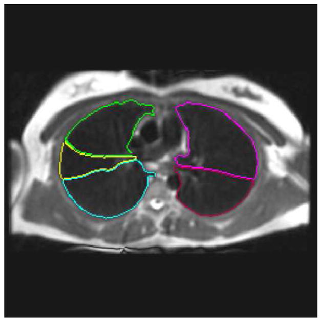
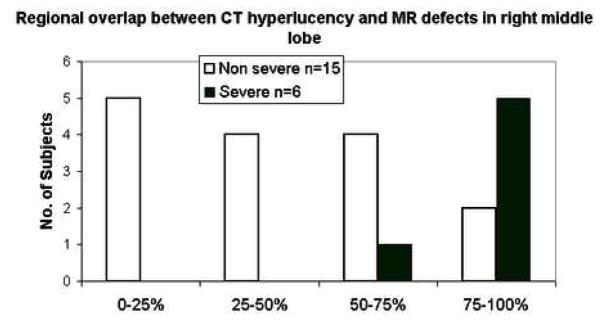
Materials and methods: Both HP MRI using helium-3 and MDCT were acquired in the same patients. Supervised segmentation of the lung lobes on MRI and MDCT facilitated regional comparisons of ventilation abnormalities in the lung parenchyma. The percentage of spatial overlap was evaluated between regions of ventilation defect on HP MRI and hyperlucency on MDCT to determine associations between obstruction and likely regions of gas trapping. Similarly, lung lobes with high defect volume were compared to lobes with low defect volume for differences in inflammatory cell number and percentage using bronchoscopic assessment.

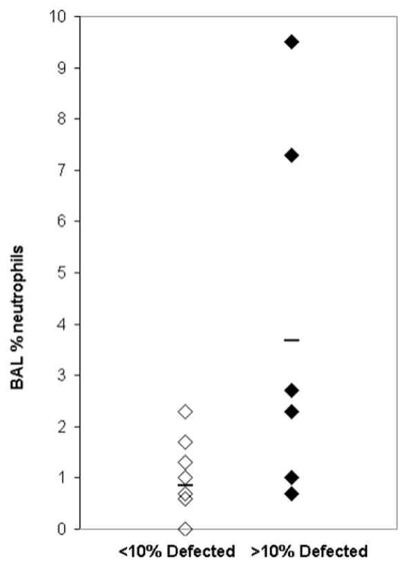
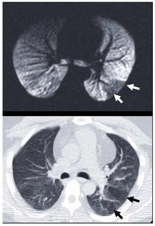
Results: There was significant overlap between sites of ventilation defect on HP MRI and hyperlucency on MDCT suggesting that sites of airway obstruction and air trapping are associated in asthma. The percent (r=0.68; P= .0039) and absolute (r=0.61; P= .0125) number of neutrophils on bronchoalveolar lavage for the sampled lung lobe also directly correlated with increased defect volume.

Conclusions: These results show promise for using image guidance to assess specific regions of ventilation defect or air trapping in heterogeneous obstructive lung diseases such as asthma.


Figures

Figure 1
Figure 1
(a) Segmented lung lobes (color code: yellow = right middle lobe, green = right upper lobe, blue = right lower lobe, magenta = left upper lobe, and red = left lower lobe) overlain on fast spin-echo (FSE) conventional proton MRI of the lung parenchyma acquired with the FSE sequence (Table 2). (b) Corresponding inspiratory MDCT slice (a) used for segmenting the lung lobes in (a). MDCT image in (b) 512 × 512, reconstructed at 0.5 cm thickness, using parameters summarized in Table 1. (c) Graphic user interface display with equivalent proton MRI and HP MRI slices with segmented lung lobes as described in (a). The HP MR image of ventilation in the right panel of (c) depicts ventilation defects segmented with lobe location marked using the same color code and was acquired with a GRE sequence and parameters summarized in Table 2.
Figure 1
Figure 1
(a) Segmented lung lobes (color code: yellow = right middle lobe, green = right upper lobe, blue = right lower lobe, magenta = left upper lobe, and red = left lower lobe) overlain on fast spin-echo (FSE) conventional proton MRI of the lung parenchyma acquired with the FSE sequence (Table 2). (b) Corresponding inspiratory MDCT slice (a) used for segmenting the lung lobes in (a). MDCT image in (b) 512 × 512, reconstructed at 0.5 cm thickness, using parameters summarized in Table 1. (c) Graphic user interface display with equivalent proton MRI and HP MRI slices with segmented lung lobes as described in (a). The HP MR image of ventilation in the right panel of (c) depicts ventilation defects segmented with lobe location marked using the same color code and was acquired with a GRE sequence and parameters summarized in Table 2.
Figure 2
Figure 2
Scatter plot of high and low defected lobes showing increased % neutrophils (p = 0.03) in lobes sampled with BAL and greater than 10% Defect Volume compared with lobes with less than 10% Defect Volume.
Figure 3
Figure 3
Typical example of the observed spatial correspondence (a) between ventilation defects on HP MRI (white arrows) and (b) hyperlucency on MDCT (black arrows). These defects in the left lower lobe reflect obstructive physiology at different lung volumes: 15% of TLC for HP MRI in (a) and FRC for expiratory MDCT in (b).
Figure 4
Figure 4
Qualitative distribution of regional overlap scores in the right middle lobe showing increased overlap between ventilation defects on HP MRI with hyperlucency on MDCT for subjects with severe vs. non-severe asthma.

References

    1. Hartert TV, Peebles RS., Jr Epidemiology of asthma: the year in review. Curr Opin Pulm Med. 2000;6(1):4–9. - PubMed
    1. Sears MR, Greene JM, Willan AR, Wiecek EM, Taylor DR, Flannery EM, Cowan JO, Herbison GP, Silva PA, Poulton R. A Longitudinal, Population-Based, Cohort Study of Childhood Asthma Followed to Adulthood. The New England Journal of Medicine. 2003;349:1414–1422. - PubMed
    1. Postma DS, Timens W. Remodeling in asthma and chronic obstructive pulmonary disease. Proc Am Thorac Soc. 2006;3(5):434–439. - PubMed
    1. Permutt S. Current status of functional pulmonary imaging. Acad Radiol. 2005;12(11):1359–1361. - PubMed
    1. Haczku A, Emami K, Fischer MC, Kadlecek S, Ishii M, Panettieri RA, Rizi RR. Hyperpolarized 3He MRI in asthma measurements of regional ventilation following allergic sensitization and challenge in mice--preliminary results. Acad Radiol. 2005;12(11):1362–1370. - PubMed