. 2008 Aug;29(23):3346-56.
doi: 10.1016/j.biomaterials.2008.04.040. Epub 2008 May 16.

An adaptable hydrogel array format for 3-dimensional cell culture and analysis

Affiliations

An adaptable hydrogel array format for 3-dimensional cell culture and analysis

Leenaporn Jongpaiboonkit et al. Biomaterials. 2008 Aug.

Abstract

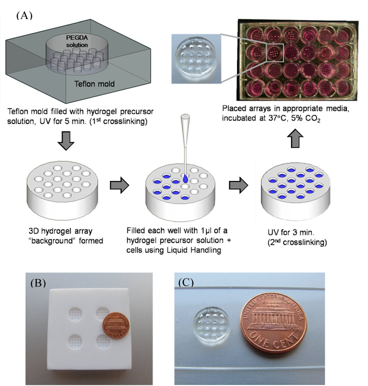
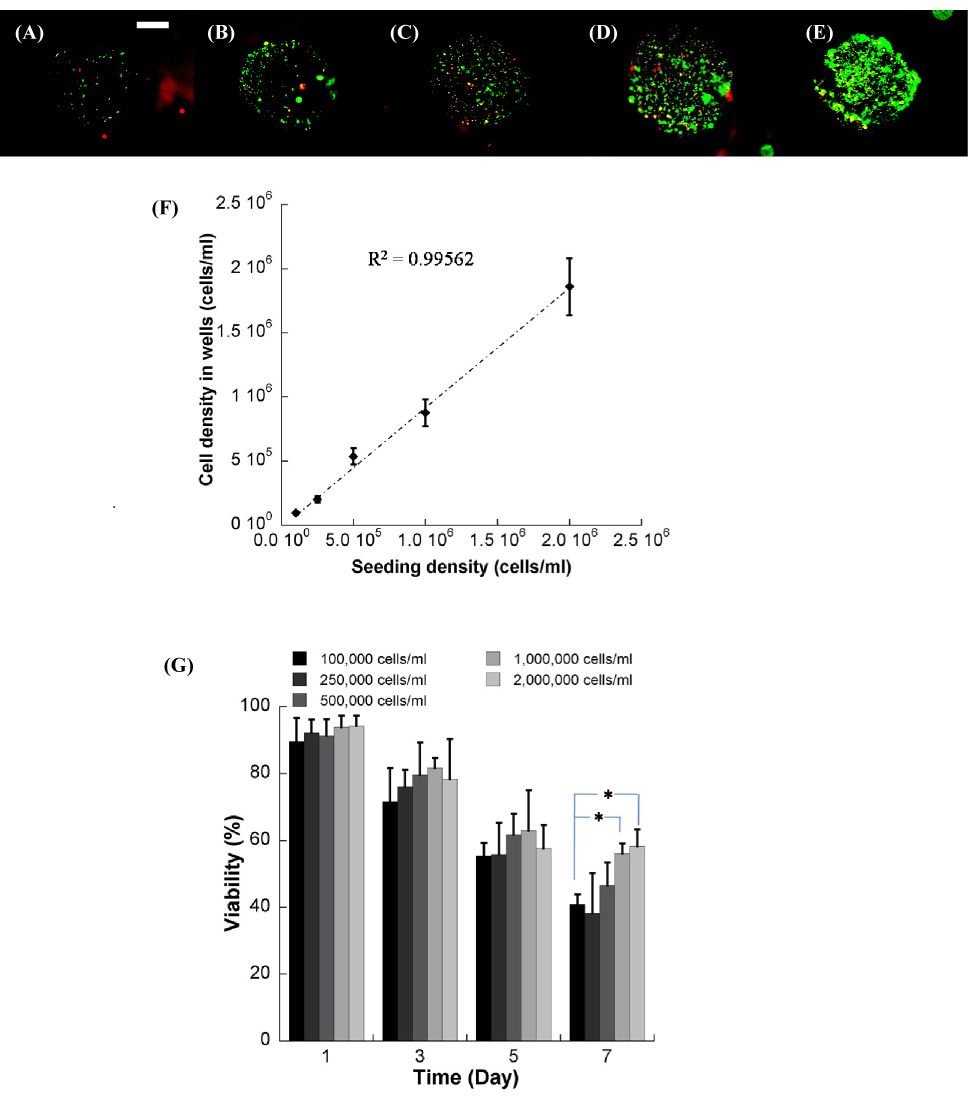
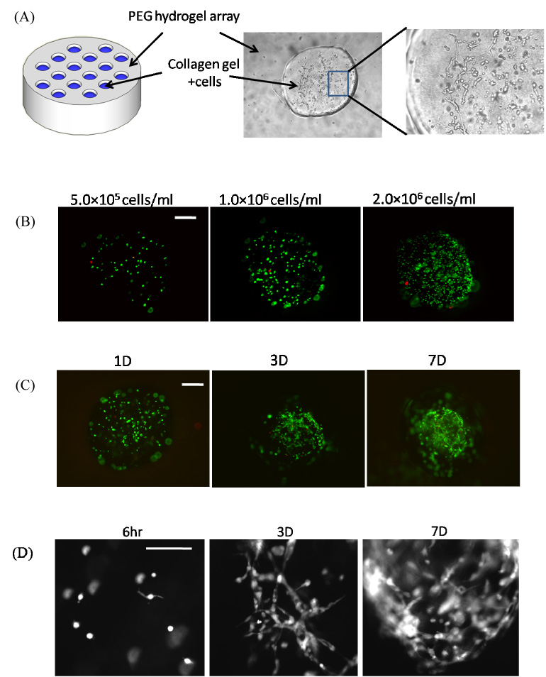
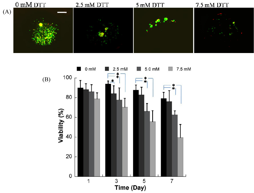
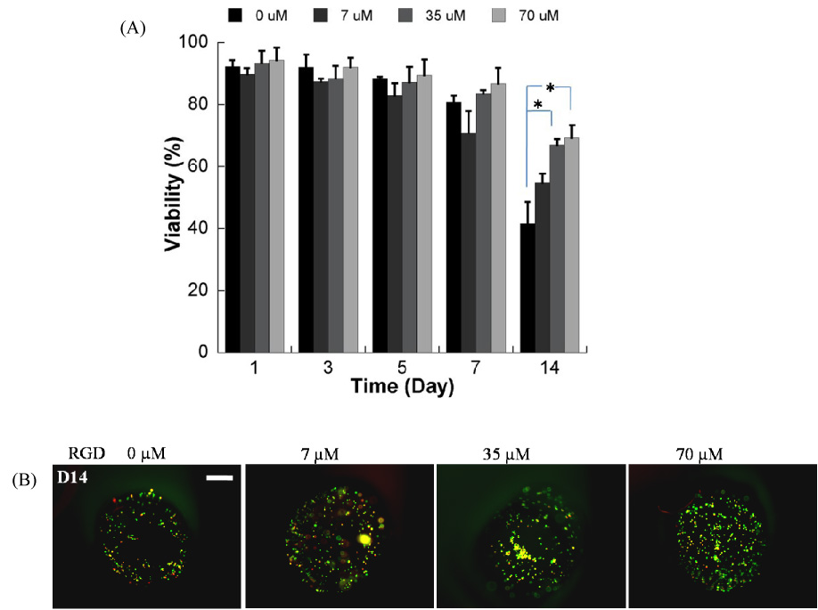
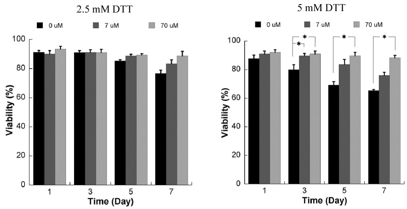
Hydrogels have been commonly used as model systems for 3-dimensional (3-D) cell biology, as they have material properties that resemble natural extracellular matrices (ECMs), and their cell-interactive properties can be readily adapted in order to address a particular hypothesis. Natural and synthetic hydrogels have been used to gain fundamental insights into virtually all aspects of cell behavior, including cell adhesion, migration, and differentiated function. However, cell responses to complex 3-D environments are difficult to adequately explore due to the large number of variables that must be controlled simultaneously. Here we describe an adaptable, automated approach for 3-D cell culture within hydrogel arrays. Our initial results demonstrate that the hydrogel network chemistry (both natural and synthetic), cell type, cell density, cell adhesion ligand density, and degradability within each array spot can be systematically varied to screen for environments that promote cell viability in a 3-D context. In a test-bed application we then demonstrate that a hydrogel array format can be used to identify environments that promote viability of HL-1 cardiomyocytes, a cell line that has not been cultured previously in 3-D hydrogel matrices. Results demonstrate that the fibronectin-derived cell adhesion ligand RGDSP improves HL-1 viability in a dose-dependent manner, and that the effect of RGDSP is particularly pronounced in degrading hydrogel arrays. Importantly, in the presence of 70mum RGDSP, HL-1 cardiomyocyte viability does not decrease even after 7 days of culture in PEG hydrogels. Taken together, our results indicate that the adaptable, array-based format developed in this study may be useful as an enhanced throughput platform for 3-D culture of a variety of cell types.


Figures

Figure 1
Figure 1
A) Schematic representation of fabrication of 3D hydrogel array. B) Teflon mold used to create hydrogel “background”. C) PEGDA 8K hydrogel array background.
Figure 2
Figure 2
Incorporation of fluorescently-labeled PEG-acrylate molecules into the background hydrogel during formation of an array spot. Fluorescein-cysteine-PEG-acrylate molecules diffuse into the hydrogel background and form a stable, interpenetrated network between the array spot and the hydrogel background.
Figure 3
Figure 3
Degradable PEG hydrogel arrays containing DTT have varying mass equilibrium swelling ratios (Qm) over time. A) Changes in Qm of PEG hydrogels with various DTT concentrations during incubation in PBS, pH=7.4, T=37°C. B) Images of hydrogel arrays prepared with different concentration of DTT at day 0 and day 6 after hydrogel preparation.
Figure 4
Figure 4
Representative images of NIH/3T3 cells incorporation into PEG hydrogel arrays A) 2 hours after encapsulation, and B) 48 hours after encapsulation. ((A) and (B) were taken at 40x magnification, scale bar = 100 µm). Representative image at higher magnification (100x magnification, scale bar = 50 µm) 2 hours after encapsulation. (D–F) Viability of three cell types in hydrogel arrays filled manually (by hand) and filled automatically (via a liquid handling robot) at 3 min UV exposure time at various time point; D) NIH/3T3 fibroblasts, E) HUVEC, and F) hMSC. (* Significant difference between manual and automated array processing methods within the same time points, p<0.05, t test).
Figure 5
Figure 5
NIH/3T3 cells seeded in the well of the hydrogel array at various cell seeding densities, (A) 1.0×10⁵, (B) 2.5×10⁵,(C) 5.0×10⁵, (D) 1.0×10⁶, and (E) 2.0×10⁶ cells/ml respectively. F) Relationship between cell density in the hydrogel precursor solution and cell density measured in the wells. G) Viability of NIH/3T3 cells seeded at various densities over the range of 7 days. * Significant difference within the same time points, p<0.05. (40x magnification, scale bar = 100 µm).
Figure 6
Figure 6
A) Schematic representation of PEG hydrogel array filled with collagen hydrogels in spots. B) Seeding of NIH/3T3 fibroblasts into collagen hydrogel spots with various cell seeding densities. C) Live/dead images of NIH/3T3 fibroblasts cultured in collagen hydrogel spots for various times. D) Higher magnification (100X) images of NIH/3T3 fibroblasts spreading over time in collagen hydrogel array spots.
Figure 7
Figure 7
A) Live/Dead images demonstrating the influence of matrix degradation on viability of HL-1 cells encapsulated in PEG hydrogel arrays with varying degradability, 7 days after hydrogel preparation (seeding density=1.0×10⁶ cells/ml) (40x magnification, scale bar = 100 µm). B) Quantitative analysis of HL-1 cardiomyocyte viability in hydrogel arrays with varying degradability. (* Significant difference between conditions with varying DTT content, within the same time points, p<0.05).
Figure 8
Figure 8
A) Quantitative analysis of HL-1 cardiomyocyte viability within non-degradable PEG hydrogel arrays containing variable RGDSP concentrations (* Significant difference within the same time points, p<0.05); B) Representative images of live/dead staining of cells in array spots 14 days after array preparation demonstrating the influence of RGDSP on HL-1 cardiomyocyte viability. (40x magnification, scale bar = 100 µm).
Figure 9
Figure 9
Influence of RGD incorporation on viability of HL-1 cells encapsulated within degradable PEG hydrogels using the live/dead assay. Data are shown for hydrogels prepared with 2.5mM (left) or 5mM DTT (right), and variable concentrations of the RGDSP peptide * Significant difference within the same time points, p<0.05.

References

    1. Edelman DB, Keefer EW. A cultural renaissance: in vitro cell biology embraces three-dimensional context. Exp Neurol. 2005;192(1):1–6. - PubMed
    1. Cukierman E, Pankov R, Yamada KM. Cell interactions with three-dimensional matrices. Curr Opin Cell Biol. 2002;14(5):633–639. - PubMed
    1. Green JA, Yamada KM. Three-dimensional microenvironments modulate fibroblast signaling responses. Adv Drug Deliv Rev. 2007;59(13):1293–1298. - PMC - PubMed
    1. Cukierman E, Pankov R, Stevens DR, Yamada KM. Taking cell-matrix adhesions to the third dimension. Science. 2001;294(5547):1708–1712. - PubMed
    1. Vaalamo M, Mattila L, Johansson N, Kariniemi AL, Karjalainen-Lindsberg ML, Kahari VM, et al. Distinct populations of stromal cells express collagenase-3 (MMP-13) and collagenase-1 (MMP-1) in chronic ulcers but not in normally healing wounds. J Invest Dermatol. 1997;109(1):96–101. - PubMed

Publication types