

Reappraisal of velocity criteria for carotid bulb/internal carotid artery stenosis utilizing high-resolution B-mode ultrasound validated with computed tomography angiography
- PMID: 18486416
- PMCID: PMC3506188
- DOI: 10.1016/j.jvs.2008.02.068
Reappraisal of velocity criteria for carotid bulb/internal carotid artery stenosis utilizing high-resolution B-mode ultrasound validated with computed tomography angiography
Abstract
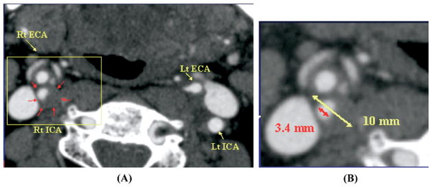
Objective: Reliability of the most commonly used duplex ultrasound (DUS) velocity thresholds for internal carotid artery (ICA) stenosis has been questioned since these thresholds were developed using less precise methods to grade stenosis severity based on angiography. In this study, maximum percent diameter carotid bulb ICA stenosis (European Carotid Surgery Trial [ECST] method) was objectively measured using high resolution B-mode DUS validated with computed tomography angiography (CTA) and used to determine optimum velocity thresholds for > or =50% and > or =80% bulb internal carotid artery stenosis (ICA).
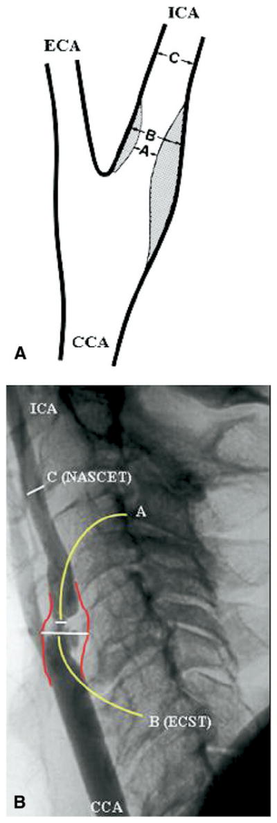
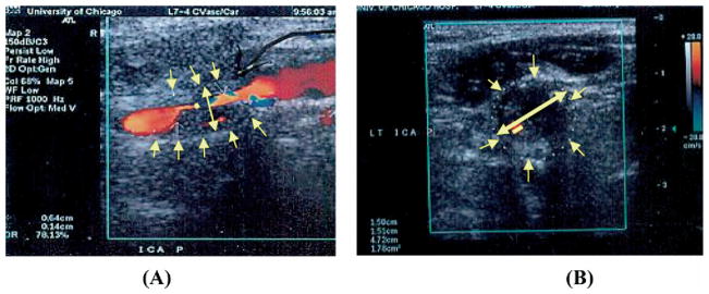
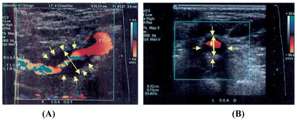
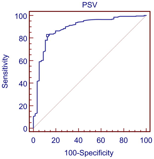
Methods: B-mode DUS and CTA images of 74 bulb ICA stenoses were compared to validate accuracy of the DUS measurements. In 337 mild, moderate, and severe bulb ICA stenoses (n = 232 patients), the minimal residual lumen and the maximum outer bulb/proximal ICA diameter were determined on longitudinal and transverse images. This in contrast to the North American Symptomatic Carotid Endarterectomy Trial (NASCET) method using normal distal ICA lumen diameter as the denominator. Severe calcified carotid segments and patients with contralateral occlusion were excluded. In each study, the highest peak systolic (PSV) and end-diastolic (EDV) velocities as well as ICA/common carotid artery (CCA) ratio were recorded. Using receiver operating characteristic (ROC) analysis, the optimum threshold for each hemodynamic parameter was determined to predict > or =50% (n = 281) and > or =80% (n = 62) bulb ICA stenosis.
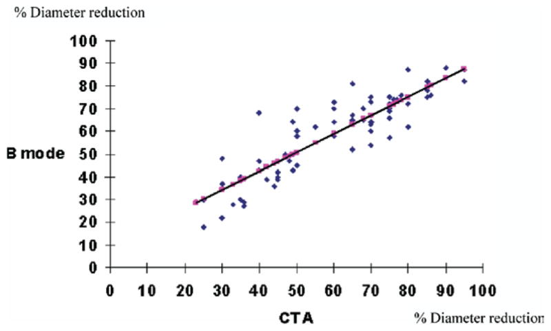
Results: Patients mean age was 74 +/- 8 years; 49% females. Clinical risk factors for atherosclerosis included coronary artery disease (40%), diabetes mellitus (32%), hypertension (70%), smoking (34%), and hypercholesterolemia (49%). Thirty-three percent of carotid lesions (n = 110) presented with ischemic cerebrovascular symptoms and 67% (n = 227) were asymptomatic. There was an excellent agreement between B-mode DUS and CTA (r = 0.9, P = .002). The inter/intraobserver agreement (kappa) for B-mode imaging measurements were 0.8 and 0.9, respectively, and for CTA measurements 0.8 and 0.9, respectively. When both PSV of > or =155 cm/s and ICA/CCA ratio of > or =2 were combined for the detection of > or =50% bulb ICA stenosis, a positive predictive value (PPV) of 97% and an accuracy of 82% were obtained. For a > or =80% bulb ICA stenosis, an EDV of > or =140 cm/s, a PSV of > or =370 cm/s and an ICA/CCA ratio of > or =6 had acceptable probability values.
Conclusion: Compared with established velocity thresholds commonly applied in practice, a substantially higher PSV (155 vs 125 cm/s) was more accurate for detecting > or =50% bulb/ICA stenosis. In combination, a PSV of > or =155 cm/s and an ICA/CCA ratio of > or =2 have excellent predictive value for this stenosis category. For > or =80% bulb ICA stenosis (NASCET 60% stenosis), an EDV of 140 cm/s, a PSV of > or =370 cm/s, and an ICA/CCA ratio of > or =6 are equally reliable and do not indicate any major change from the established criteria. Current DUS > or =50% bulb ICA stenosis criteria appear to overestimate carotid bifurcation disease and may predispose patients with asymptomatic carotid disease to untoward costly diagnostic imaging and intervention.
Conflict of interest statement
Competition of interest: none.
Figures
References
-
- North American Symptomatic Carotid Endarterectomy Trial Collaborators. Beneficial effect of carotid endarterectomy in symptomatic patients with high-grade carotid stenosis. N Engl J Med. 1991;325:445–53. - PubMed
-
- European Carotid Surgery Trialists’ Collaborative Group. Randomized trial of endarterectomy for recently symptomatic carotid stenosis: final results of the MRC European Carotid Surgery Trial (ECST) Lancet. 1998;351:1379–87. - PubMed
-
- Endarterectomy for asymptomatic carotid artery stenosis. Executive Committee for the Asymptomatic Carotid Atherosclerosis Study. JAMA. 1995;273:1421–8. - PubMed
-
- Blakeley DD, Oddone EZ, Hasselblad V, Simel DL, Matchar DB. Noninvasive carotid artery testing: a meta-analytic review. Ann Intern Med. 1995;122:360–7. - PubMed
-
- Zierler RE, Strandness DE., Jr . Noninvasive dynamic and real-time assessment of extracranial cerebrovasculature. In: Wood JH, editor. Cerebral blood flow: physiologic and clinical aspects. New York: McGraw Hill; 1987. pp. 311–23.
MeSH terms
Grants and funding
LinkOut - more resources
Full Text Sources
Miscellaneous
