

Evaluation of surgically retrieved temporomandibular joint alloplastic implants: pilot study
- PMID: 18486775
- PMCID: PMC2670201
- DOI: 10.1016/j.joms.2007.09.013
Evaluation of surgically retrieved temporomandibular joint alloplastic implants: pilot study
Abstract
Purpose: The purpose of this study was to perform a retrieval analysis of temporomandibular joint (TMJ) alloplastic interpositional implants and test possible correlation between implant failure features and patient clinical outcomes. In addition, we investigated the implants' surface and examined the foreign body reaction associated with different types of alloplastic materials.
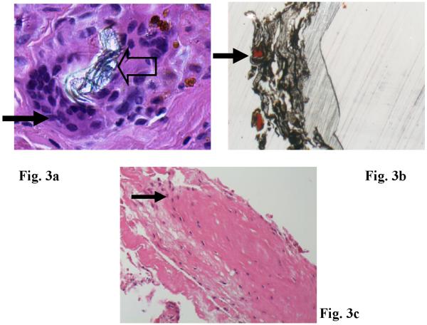
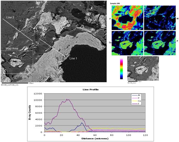
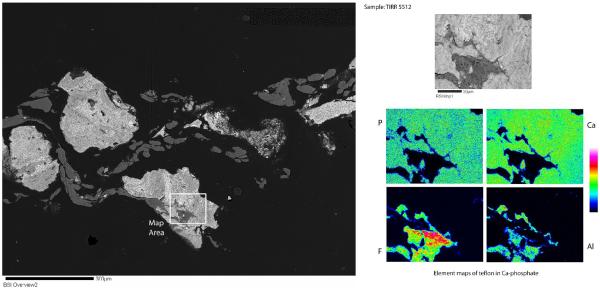
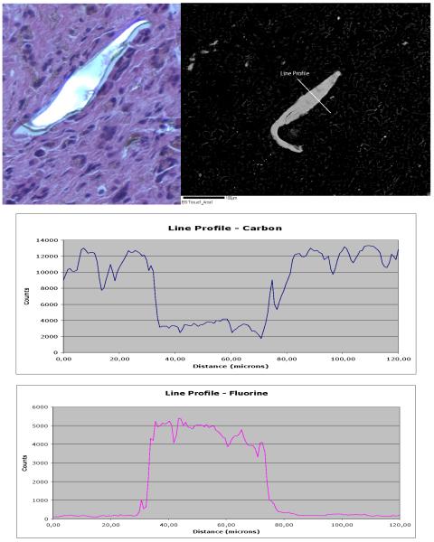
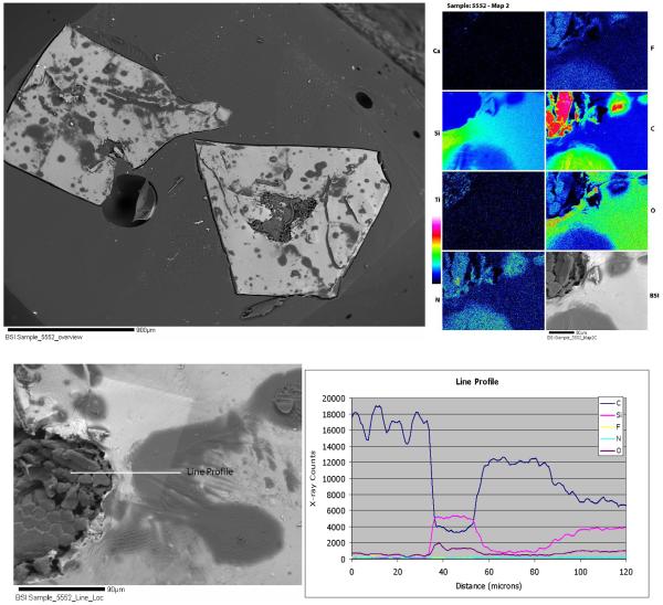
Materials and methods: Twelve implants (Proplast/Teflon [Vitek, Houston, TX] and Silastic [Dow Corning, Midland, MI]) were surgically removed from the patients' TMJs. Implant surface failure features (fracture length, perforation of the implants) were observed using stereomicroscopy and recorded for description of the failure mechanisms and to statistically compare with clinical outcomes. Patients' clinical data (pain symptoms and mandibular function) were collected and examined. Clinical outcomes were obtained relative to symptom severity (Symptom Severity Index [SSI]) and jaw function (modified Mandibular Function Impairment Questionnaire [mMFIQ]). Peri-implant soft tissues and implants were analyzed with light microscopy and stereo zoom microscopy. Electron microprobe analysis of implant fragments and peri-implant tissues was performed.
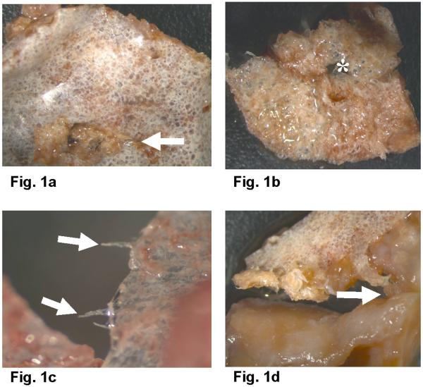
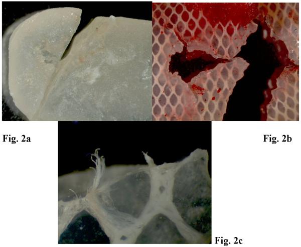
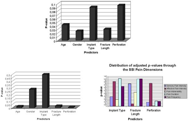
Results: The statistical results showed that only the presence of implant perforation was statistically associated with the SSI, specifically with the pain tolerability dimension. No statistical association was seen between any of the other implant failure predictors and the SSI and between the predictors and the mMFIQ. Stereo zoom microscopy suggested that Proplast/Teflon implants (n = 7) were susceptible to perforation, layer tearing, fracture and fiber extrusion. The Silastic implants (n = 3) revealed a possible center perforation with fracture lines towards the periphery and fiber extrusion. Teflon implant wear debris particles appear to trigger a multinucleated giant cell foreign body reaction.
Conclusion: Facial pain was a significant correlate to perforation and breakdown of the alloplastic TMJ interpositional implants, and most likely was the reason for implant removal.
Figures
References
-
- Okeson JP. Orofacial pain: guidelines for assessment, diagnosis, and management, The American Academy of Orofacial Pain. Quintessence Publishing Co Inc.; Carol Stream, IL: 1996. pp. 113–158.
-
- Okeson JP. Management of Temporomandibular disorders and Occlusion. 5th edition Mosby Inc.; 2003. pp. 3–27.
-
- Okeson JP. Bell's Orofacial Pains: The Clinical Management of Orofacial Pain. 6th edition Quintessence Publishing Co Inc.; Carol Stream, IL: 2005. pp. 287–373.
-
- Wright EF. Manual of Temporomandibular disorders. Blackwell Munksgaard; Ames, IO: 2005. pp. 181–256.
-
- Dimitroulis G. The role of surgery in the management of disorders of the Temporomandibular Joint: a critical review of the literature. Int J Oral Maxillofac Surg. 2005;34(Part 1):107–113. - PubMed
Publication types
MeSH terms
Substances
Grants and funding
LinkOut - more resources
Full Text Sources
Medical
