

Mucus secretion and cytoskeletal modifications in cultured nasal epithelial cells exposed to wall shear stresses
- PMID: 18487304
- PMCID: PMC2527286
- DOI: 10.1529/biophysj.107.127142
Mucus secretion and cytoskeletal modifications in cultured nasal epithelial cells exposed to wall shear stresses
Abstract
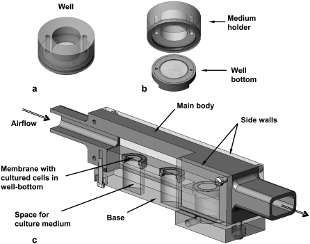
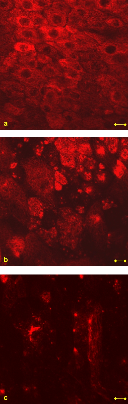
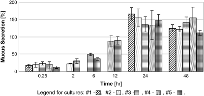
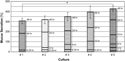
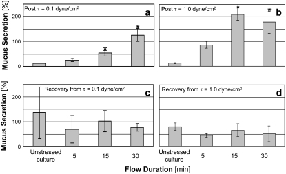
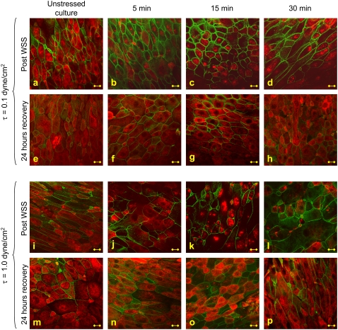
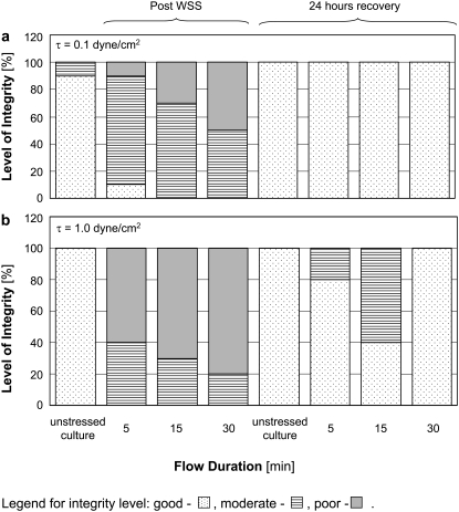
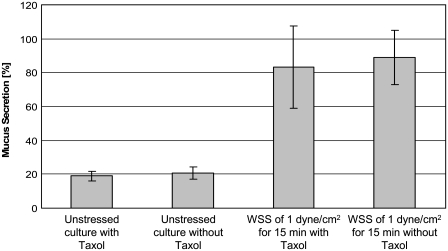
The nasal epithelium is continuously subjected to wall shear stresses (WSS) induced by respiratory airflows. An in vitro experimental model was developed to expose nasal epithelial cells cultured under air-liquid interface conditions to steady airflow-induced WSS. Mucus secretion from epithelial goblet cells was quantified using an enzyme-linked lectinosorbent assay, and modifications of the cytoskeletal structure were qualitatively evaluated from fluorescent stains of actin and beta-tubulin fibers. The results show increased mucus secretion from cells subjected to WSS of 0.1 and 1.0 dyne/cm(2) for more than 15 min in comparison with unstressed cells. The integrity levels of beta-tubulin fibers were significantly lower in cells subjected to WSS than in unstressed cells. The increased mucus secretion in response to WSS was approximately the same in Taxol-free and Taxol-treated cultures, which indicates that there is no direct connection between beta-tubulin fragmentation and mucus secretion. The stressed cells regained their normal cytoskeletal appearance 24 h after the exposure to WSS. The results of this study suggest that WSS have an important role in the mechanical regulation of the nasal surface epithelium function.
Figures
Similar articles
-
Mechanobiology of conjunctival epithelial cells exposed to wall shear stresses.Biomech Model Mechanobiol. 2021 Oct;20(5):1903-1917. doi: 10.1007/s10237-021-01484-y. Epub 2021 Jul 6. Biomech Model Mechanobiol. 2021. PMID: 34228228
-
Mechanophysical stimulations of mucin secretion in cultures of nasal epithelial cells.Biophys J. 2011 Jun 22;100(12):2855-64. doi: 10.1016/j.bpj.2011.04.040. Biophys J. 2011. PMID: 21689518 Free PMC article.
-
A nasal airway-on-chip model to evaluate airflow pre-conditioning during epithelial cell maturation at the air-liquid interface.Biofabrication. 2024 Aug 13;16(4). doi: 10.1088/1758-5090/ad663d. Biofabrication. 2024. PMID: 39038483
-
Transmembrane Mucin Response in Conjunctival Epithelial Cells Exposed to Wall Shear Stresses.Int J Mol Sci. 2023 Apr 1;24(7):6589. doi: 10.3390/ijms24076589. Int J Mol Sci. 2023. PMID: 37047561 Free PMC article.
-
Epithelial mucus-hypersecretion and respiratory disease.Curr Drug Targets Inflamm Allergy. 2005 Dec;4(6):651-64. doi: 10.2174/156801005774912851. Curr Drug Targets Inflamm Allergy. 2005. PMID: 17305521 Review.
Cited by
-
[Numerical flow simulation : A new method for assessing nasal breathing].HNO. 2016 Aug;64(8):611-8. doi: 10.1007/s00106-016-0209-8. HNO. 2016. PMID: 27455988 German.
-
Proteomics of buccal cavity mucus in female tilapia fish (Oreochromis spp.): a comparison between parental and non-parental fish.PLoS One. 2011 Apr 20;6(4):e18555. doi: 10.1371/journal.pone.0018555. PLoS One. 2011. PMID: 21533134 Free PMC article.
-
Fluid-flow induced wall shear stress and epithelial ovarian cancer peritoneal spreading.PLoS One. 2013 Apr 10;8(4):e60965. doi: 10.1371/journal.pone.0060965. Print 2013. PLoS One. 2013. PMID: 23593358 Free PMC article.
-
Numerical modelling of micron particle inhalation in a realistic nasal airway with pediatric adenoid hypertrophy: A virtual comparison between pre- and postoperative models.Front Pediatr. 2023 Feb 23;11:1083699. doi: 10.3389/fped.2023.1083699. eCollection 2023. Front Pediatr. 2023. PMID: 36911037 Free PMC article.
-
The Path from Nasal Tissue to Nasal Mucosa on Chip: Part 1-Establishing a Nasal In Vitro Model for Drug Delivery Testing Based on a Novel Cell Line.Pharmaceutics. 2023 Aug 30;15(9):2245. doi: 10.3390/pharmaceutics15092245. Pharmaceutics. 2023. PMID: 37765214 Free PMC article.
References
-
- Kim, K. C., J. Nassiri, and J. S. Brody. 1989. Mechanisms of airway goblet cell mucin release: studies with cultured tracheal surface epithelial cells. Am. J. Respir. Cell Mol. Biol. 1:137–143. - PubMed
-
- Kim, K. C. 1991. Biochemistry and pharmacology of mucin-like glycoproteins produced by cultured airway epithelial cells. Exp. Lung Res. 17:533–545. - PubMed
-
- Rogers, D. F. 1994. Airway goblet cells: responsive and adaptable front-line defenders. Eur. Respir. J. 7:1690–1708. - PubMed
MeSH terms
LinkOut - more resources
Full Text Sources
Research Materials
