Review
. 2008 Jul;95(2):497-502.
doi: 10.1529/biophysj.108.131797. Epub 2008 May 16.

Packaging double-helical DNA into viral capsids: structures, forces, and energetics

Affiliations
Review

Packaging double-helical DNA into viral capsids: structures, forces, and energetics

Anton S Petrov et al. Biophys J. 2008 Jul.

Abstract

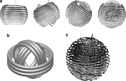
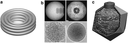
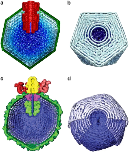
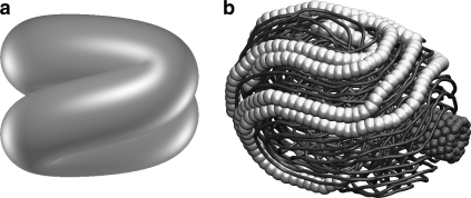
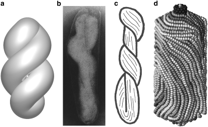
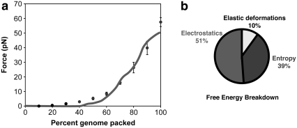
Small, icosahedral double-stranded DNA bacteriophage pack their genomes tightly into preformed protein capsids using an ATP-driven motor. Coarse-grain molecular-mechanics models provide a detailed picture of DNA packaging in bacteriophage, revealing how conformation depends on capsid size and shape, and the presence or absence of a protein core. The forces that oppose packaging have large contributions from both electrostatic repulsions and the entropic penalty of confining the DNA into the capsid, whereas elastic deformations make only a modest contribution. The elastic deformation energy is very sensitive to the final conformation, whereas the electrostatic and entropic penalties are not, so the packaged DNA favors conformations that minimize the bending energy.


Figures

FIGURE 1
FIGURE 1
Concentric spools. (a) Sequence of energy-minimized structures for DNA packed into a spherical capsid with no core (26). (b) Idealized representation. (c) DNA packaged in a spherical capsid with no core at T = 300 K (35).
FIGURE 2
FIGURE 2
Coaxial spools. (a) Idealized representation. (b) Simulated (32) (upper two panels) and experimental (40) (lower two panels) transmission EM density maps for T7 DNA. Projections of single particles along an axis perpendicular to the axis of packaging give a punctate pattern (left two panels), whereas projections along the axis of packaging give concentric rings when multiple images are averaged (right two panels): 10 independent packaging trajectories for the simulations, and 70 independent particles for the experiments. (c) Cutaway view of a single coaxially spooled conformation for a model of ɛ15 (34). To permit a clearer view of the DNA organization, the graphical diameter of the DNA strands is ∼8 Å (much less than the DNA-DNA contact distance of 25 Å).
FIGURE 3
FIGURE 3
Single-particle reconstructions of electron density maps for ɛ15 from simulations (34) (panels a and b) and experiment (41) (panels c and d). The complete capsid and core are shown in the transverse cutaway views (a and c), whereas only the DNA density is shown in the views down the packaging axis (b and d). The central closed circular ring of density in views b and d is a consequence of averaging over many conformations (40 independent packaging trajectories in the simulations, and ∼15,000 particles in the experimental reconstruction). The ring of density is due to the groove in the core protein, which, from very early in the packaging trajectory, has high DNA density. Build-up of pressure in this groove is believed to drive a conformational switch in the portal protein, signaling that the capsid is full (43).
FIGURE 4
FIGURE 4
Folded toroidal conformation. (a) Idealized structure proposed by Hud (7). (b) Simulation of DNA packed into the slightly elongated capsid of φ29, which has a very short core (33). Individual pseudoatoms (6 bp) are shown in some regions to facilitate recognition of the characteristic pattern, which resembles the seams on a baseball.
FIGURE 5
FIGURE 5
Twisted toroidal conformation. (a) Idealized structure. (b) Electron micrograph of a partially disrupted giant T4 phage (4), which led to (c) the original model of Earnshaw et al. (4). (d) Simulation of 39.7 kbp of DNA packed into an elongated icosahedral capsid with an axial ratio of ∼3:1 (35).
FIGURE 6
FIGURE 6
Energetics of packaging. (a) Force versus distance curve for the packaging of DNA into φ29, as determined in single-molecule pulling experiments (red line) (8), and as measured in a simulation (points) (33). Error bars are ±1 SD. (b) About 39% of the free-energy cost of packaging DNA into the viral capsid is due to the entropic penalty of confinement. The remainder of the cost is due to the change in internal energy, including electrostatic repulsions (51%) and elastic bending deformations (10%). The contributions from DNA stretching, DNA-DNA hard-core repulsions (volume exclusion), and DNA-capsid volume exclusion repulsions are all negligible.

Similar articles

Cited by

References

    1. Richards, K. E., R. C. Williams, and R. Calendar. 1973. Mode of DNA packing within bacteriophage heads. J. Mol. Biol. 78:255–259. - PubMed
    1. Black, L. W., and D. J. Silverman. 1978. Model for DNA packaging into bacteriophage T4 heads. J. Virol. 28:643–655. - PMC - PubMed
    1. Hall, S. B., and J. A. Schellman. 1982. Flow dichroism of capsid DNA phages. II. Effect of DNA deletions and intercalating dyes. Biopolymers. 21:2011–2031. - PubMed
    1. Earnshaw, W. C., J. King, S. C. Harrison, and F. A. Eiserling. 1978. The structural organization of DNA packaged within the heads of T4 wild-type, isometric and giant bacteriophages. Cell. 14:559–568. - PubMed
    1. Earnshaw, W. C., and S. C. Harrison. 1977. DNA arrangement in isometric phage heads. Nature. 268:598–602. - PubMed

Publication types

LinkOut - more resources