. 2008 Jul;190(14):4971-8.
doi: 10.1128/JB.01849-07. Epub 2008 May 16.

A cellulose synthase-like protein involved in hyphal tip growth and morphological differentiation in streptomyces

Affiliations

A cellulose synthase-like protein involved in hyphal tip growth and morphological differentiation in streptomyces

Hongbin Xu et al. J Bacteriol. 2008 Jul.

Abstract

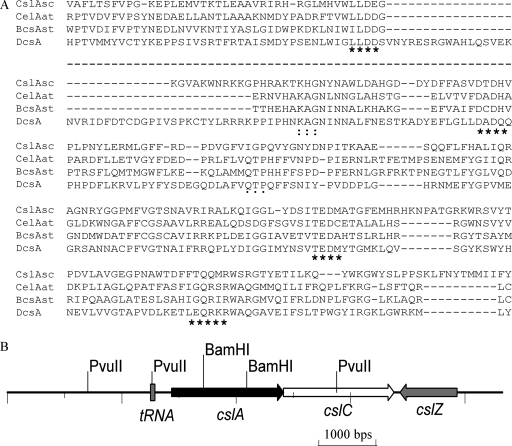
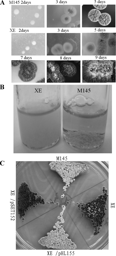
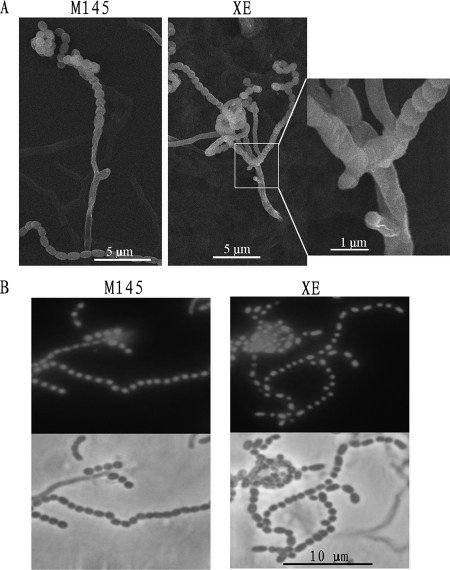
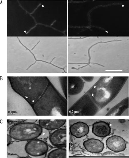
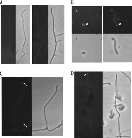
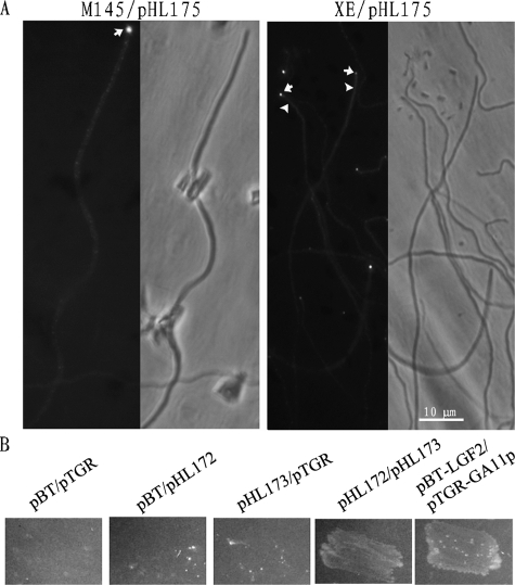
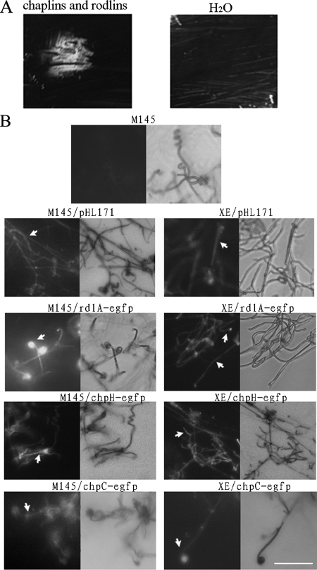
Cellulose synthase and cellulose synthase-like proteins, responsible for synthesizing beta-glucan-containing polysaccharides, play a fundamental role in cellular architectures, such as plant cell and tissue morphogenesis, bacterial biofilm formation, and fruiting-body development. However, the roles of the proteins involved in the developmental process are not well understood. Here, we report that a cellulose synthase-like protein (CslA(Sc)) in Streptomyces has a function in hyphal tip growth and morphological differentiation. The cslA(Sc) replacement mutant showed pleiotropic defects, including the severe delay of aerial-hyphal formation and altered cell wall morphology. Calcofluor white fluorescence analysis demonstrated that polysaccharide synthesis at hyphal tips was dependent on CslA(Sc). cslA(Sc) was constitutively transcribed, and an enhanced green fluorescent protein-CslA(Sc) fusion protein was mostly located at the hyphal tips. An extract enriched in morphogenetic chaplin proteins promoted formation of aerial hyphae by the mutant. Furthermore, a two-hybrid experiment indicated that the glycosyltransferase domain of CslA(Sc) interacted with the tropomyosin-like polarity-determining DivIVA protein, suggesting that the tip-located DivIVA governed tip recruitment of the CslA(Sc) membrane protein. These results imply that the cellulose synthase-like protein couples extracellular and cytoskeletal components functioning in tip growth and cell development.


Figures

FIG. 1.
FIG. 1.
The cslASc gene and comparison of its product with related proteins. (A) Alignment of the highly conserved U1, U2, U3, and U4 regions in the central cytoplasmic loop of the predicted S. coelicolor cellulose synthase-like protein with cellulose synthase protein sequences from Agrobacterium tumefaciens (CelAAt; NP_533806), S. enterica serovar Typhimurium (BcsA; CAC86199), and Dictyostelium discoideum (DcsA; AAF00200). The D, D, and D35QXXRW motifs; KAG motif; and QTP motif are indicated by asterisks, colons, and dots, respectively. The dashed line between the first and second blocks indicates that some regions that do not contain conserved motifs are omitted. (B) The csl genes in S. coelicolor. Compared with the operon of S. enterica serovar Typhimurium/E. coli, Streptomyces does not have the bcsB gene. The csl genes also exist in S. avermitilis.
FIG. 2.
FIG. 2.
Disrupting cslASc in S. coelicolor. (A) Aerial-mycelium formation by the mutant was severely delayed on solid R2YE medium compared with that of the wild-type M145. (B) Clumping, and resulting sedimentation, of vegetative hyphae in liquid culture (TSB; 24 h) was less pronounced in the cslA mutant than in the wild-type M145. Mycelial clumps of M145 sedimented to the bottom quickly when the bottle was allowed to stand (right). (C) Genetic complementation of the cslA mutant. The mutant containing pHL155 formed normal aerial hyphae on solid R2YE medium, but the mutant containing the empty plasmid pSET152 did not. The wild-type M145, XE, and XE/pSET152 were used as controls.
FIG. 3.
FIG. 3.
The cslASc gene affected aerial-hyphal development. (A) Multiple closely spaced sporulating hyphae of the mutant often emerged from one supporting hypha. Usually, the sporulating hyphae of wild-type M145 were well separated. (B) Spores of the mutant and wild-type M145 stained with PI. Spores of sporulating hyphae of the mutant contained DNA with the same appearance as that of sporulating wild-type M145. DNA-free spores of the mutant were rare (R2YE solid medium; analysis of the mutant was delayed compared to the wild type until spore chains could be detected).
FIG. 4.
FIG. 4.
Calcofluor white staining and TEM analysis. (A) Calcofluor white staining revealed that β-1,4-linked polysaccharides accumulated at the tips of vegetative hyphae in the wild type (top left, arrows) but not in the XE mutant (top right). The lower panels show the equivalent phase-contrast images. The strains were cultured on MS solid medium. (B and C) Comparison by TEM of ultrathin sections of wild-type M145 and mutant XE (R2YE solid medium). Wild-type M145 showed normal and classic vegetative hyphal cell wall and septum appearance, with an electron-dense inner layer, while the mutant lacked such layers in its cell wall and septa (B, arrows). The mutant showed abnormal wrinkled spore walls (C, arrows).
FIG. 5.
FIG. 5.
Localization of CslASc-EGFP fusion protein. The CslASc-EGFP fusion protein was localized at hyphal tips. (A) Controls; aerial hypha (left) and vegetative hyphae (right) of wild-type M145 without an egfp fusion. (B, C, and D) M145/pHL179. (B) Left, spore germination; right, germ tube elongation. (C) Vegetative hyphae. (D) Aerial hyphae. (A, C, and D) Left, fluorescent images; right, phase-contrast images. (B) Top, fluorescent images; bottom, phase-contrast images. The arrows indicate the fluorescent foci at tips. The strains were grown on MS medium. Bar, 10 μm.
FIG. 6.
FIG. 6.
Interaction between DivIVASc and CslASc. (A) Localization of DivIVASc-EGFP protein in wild-type M145 and XE. The DivIVASc-EGFP protein was mostly at hyphal tips, with some smaller foci along the hyphae. In the XE mutant, the foci were also mostly at the tips (arrows), but in some aerial hyphae, the fluorescence was dispersed through the cell (arrowheads). (B) A bacterial two-hybrid experiment showed that DivIVA interacted with the glycosyltransferase domain of the CslASc protein. The reporter strain XL1-Blue MRF′ with different plasmid pairs was grown on double-selective indicator plates containing 3-AT and streptomycin. Experimental construct, pHL172/pHL173; positive control, pBT-LGF2/pRGT-GAL11P; negative controls, pBT/pRGT, pHL173/pTRG, and pBT/pHL172.
FIG. 7.
FIG. 7.
Extracellular complementation of the XE mutant by a chaplin/rodlin extract and analysis of the expression of chaplin and rodlin hydrophobins in XE by EGFP fusions. (A) Extracellular chaplins and rodlins promoted the XE mutant to form aerial mycelium. The extracted chaplin and rodlin material or control material (water) was spotted on the lawns after 1 day of mycelial growth on R2YE solid medium. The extract was seen to promote the mutant to form aerial hyphae 1.5 days after addition. Water had no effect. The photographs were captured with a Fuji FinePix S602 2 days after addition. (B) EGFP fluorescence expressed from pHL171 derivatives carrying the P1ram-egfp, rdlA-egfp, chpH-egfp, and chpC-egfp fusions in aerial hyphae of both the wild type and the XE mutant. EGFP expression was observed in aerial hyphae of both strains containing the P1ram-egfp, rdlA-egfp, chpH-egfp, and chpC-egfp reporters. The spores were inoculated on solid R2YE medium, and aerial hyphae attached to the surfaces of coverslips were analyzed 2 days after inoculation in the cases of M145 and its derivatives and after 7 days in the cases of XE and its derivatives. Wild-type M145 without EGFP was set up as the control. Bars, 10 μm.

References

    1. Bentley, S. D., K. F. Chater, A. M. Cerdeno-Tarraga, G. L. Challis, N. R. Thomson, K. D. James, D. E. Harris, M. A. Quail, H. Kieser, D. Harper, A. Bateman, S. Brown, G. Chandra, C. W. Chen, M. Collins, A. Cronin, A. Fraser, A. Goble, J. Hidalgo, T. Hornsby, S. Howarth, C. H. Huang, T. Kieser, L. Larke, L. Murphy, K. Oliver, S. O'Neil, E. Rabbinowitsch, M. A. Rajandream, K. Rutherford, S. Rutter, K. Seeger, D. Saunders, S. Sharp, R. Squares, S. Squares, K. Taylor, T. Warren, A. Wietzorrek, J. Woodward, B. G. Barrell, J. Parkhill, and D. A. Hopwood. 2002. Complete genome sequence of the model actinomycete Streptomyces coelicolor A3(2). Nature 417141-147. - PubMed
    1. Bierman, M., R. Logan, K. O'Brien, E. T. Seno, R. Nagaraja Rao, and B. E. Schoner. 1992. Plasmid cloning vectors for the conjugal transfer of DNA from Escherichia coli to Streptomyces spp. Gene 11643-49. - PubMed
    1. Blondelet-Rouault, M.-H., J. Weiser, A. Lebrihi, P. Branny, and J.-L. Pernodet. 1997. Antibiotic resistance gene cassettes derived from the [pi] interposon for use in E. coli and Streptomyces. Gene 190315-317. - PubMed
    1. Campbell, J. A., G. J. Davies, V. Bulone, and B. Henrissat. 1997. A classification of nucleotide-diphospho-sugar glycosyltransferases based on amino acid sequence similarities. Biochem. J. 326929-939. - PMC - PubMed
    1. Chater, K. F. 1993. Genetics of differentiation in Streptomyces. Annu. Rev. Microbiol. 47685-713. - PubMed

Publication types

MeSH terms

LinkOut - more resources