. 2008 Sep;49(9):3747-57.
doi: 10.1167/iovs.07-1493. Epub 2008 May 16.

Peripheral refraction in normal infant rhesus monkeys

Affiliations

Peripheral refraction in normal infant rhesus monkeys

Li-Fang Hung et al. Invest Ophthalmol Vis Sci. 2008 Sep.

Abstract

Purpose: To characterize peripheral refractions in infant monkeys.

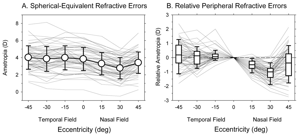
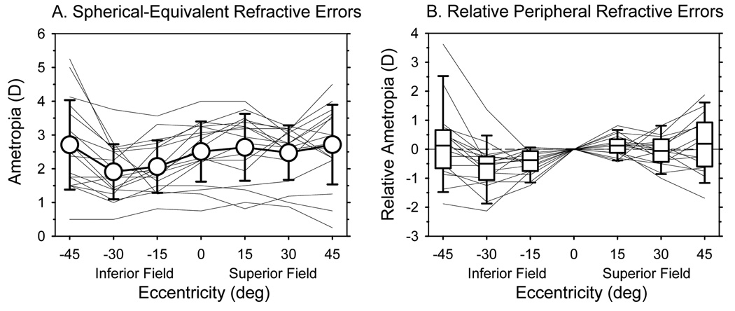
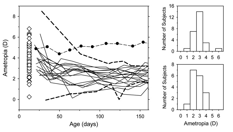
Methods: Cross-sectional data for horizontal refractions were obtained from 58 normal rhesus monkeys at 3 weeks of age. Longitudinal data were obtained for both the vertical and horizontal meridians from 17 monkeys. Refractive errors were measured by retinoscopy along the pupillary axis and at eccentricities of 15 degrees , 30 degrees , and 45 degrees . Axial dimensions and corneal power were measured by ultrasonography and keratometry, respectively.

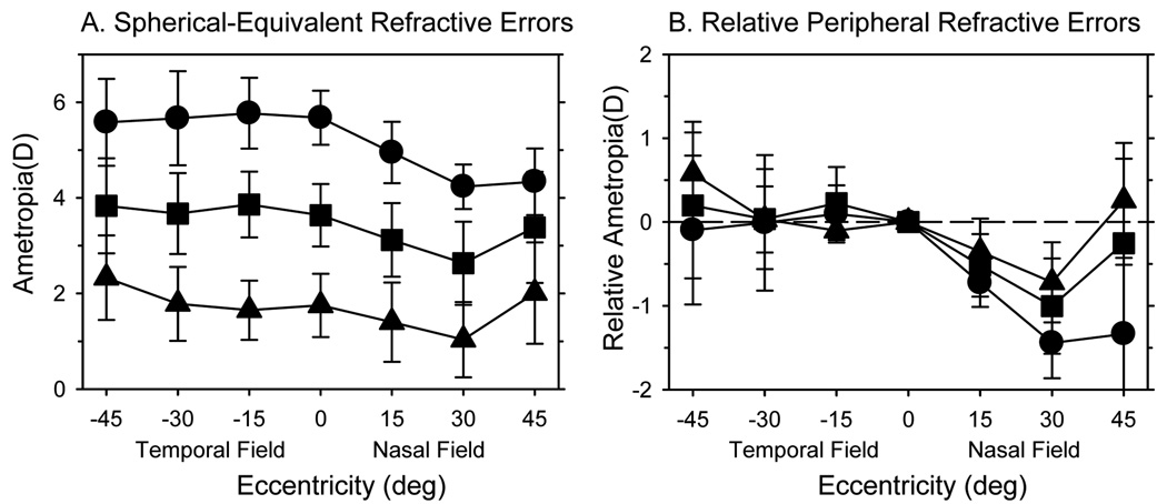
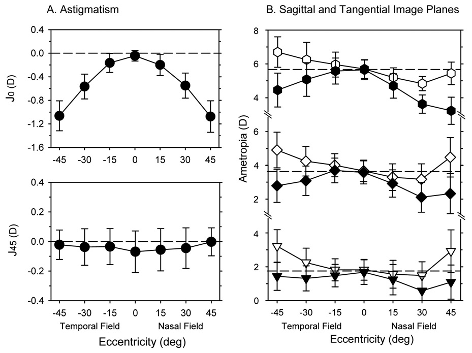
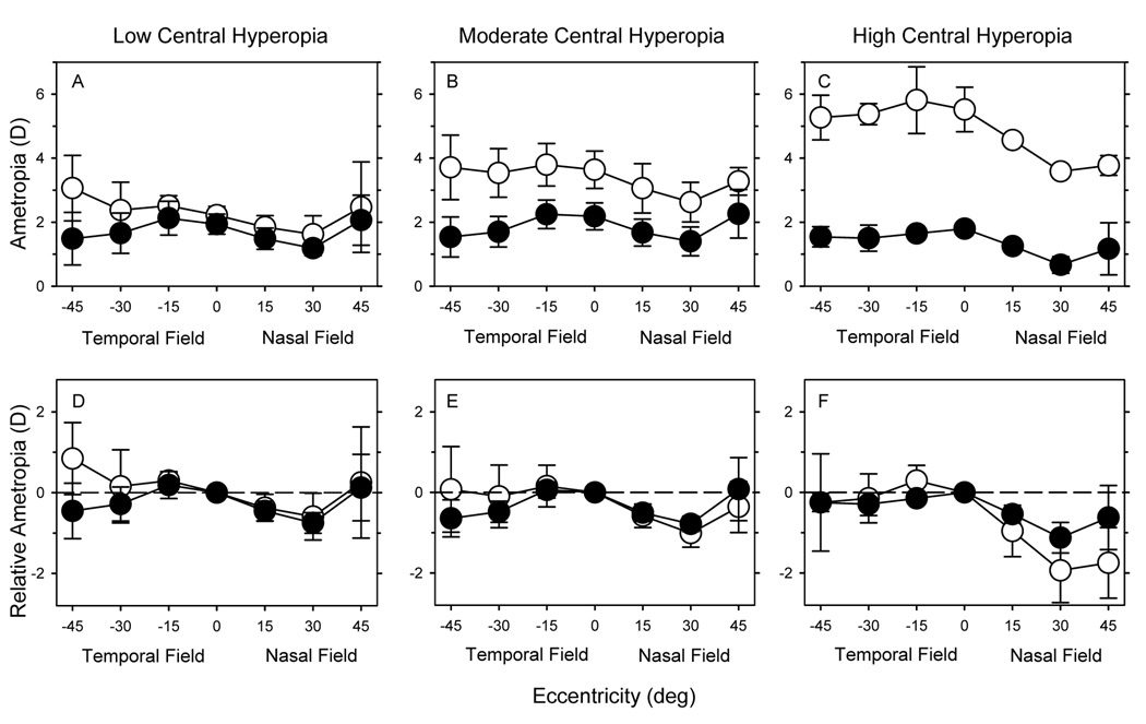
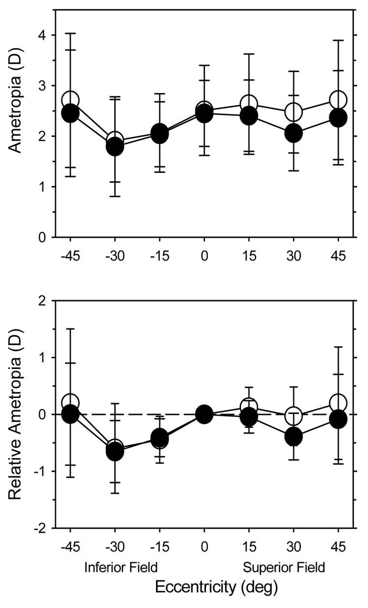
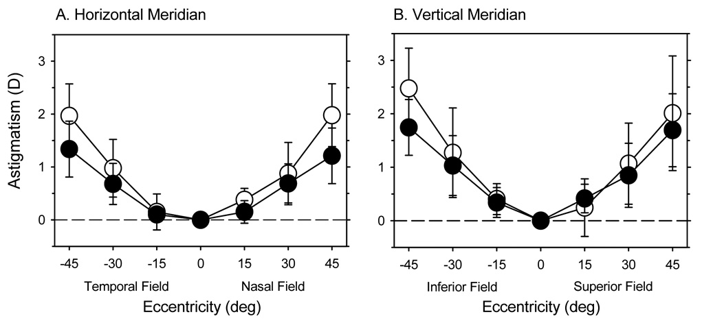
Results: In infant monkeys, the degree of radial astigmatism increased symmetrically with eccentricity in all meridians. There were, however, initial nasal-temporal and superior-inferior asymmetries in the spherical equivalent refractive errors. Specifically, the refractions in the temporal and superior fields were similar to the central ametropia, but the refractions in the nasal and inferior fields were more myopic than the central ametropia, and the relative nasal field myopia increased with the degree of central hyperopia. With age, the degree of radial astigmatism decreased in all meridians, and the refractions became more symmetrical along both the horizontal and vertical meridians. Small degrees of relative myopia were evident in all fields.

Conclusions: As in adult humans, refractive error varied as a function of eccentricity in infant monkeys and the pattern of peripheral refraction varied with the central refractive error. With age, emmetropization occurred for both central and peripheral refractive errors, resulting in similar refractions across the central 45 degrees of the visual field, which may reflect the actions of vision-dependent, growth-control mechanisms operating over a wide area of the posterior globe.


Figures

Figure 1
Figure 1
A. Spherical-equivalent refractive errors plotted as a function of horizontal eccentricity. Thin solid lines represent the right eyes of individual monkeys. The open circles represent the averages (± SD) for all 58 monkeys. B. The peripheral refractions of individual monkeys normalized to their central refractive errors plotted as a function of horizontal eccentricity. Thin solid lines represent individual monkeys. The lines in the box plots represent the medians. The boundaries of the box plots indicate the 25th and the 75th percentiles. The error bars above and below the boxes indicate the 90th and 10th percentiles, respectively.
Figure 2
Figure 2
A. Average spherical-equivalent refractive errors (± SD) plotted as a function of horizontal eccentricity for the right eyes of the monkeys in the high (filled circles), moderate (filled squares) and low hyperopic subgroups (filled triangles). B. Average peripheral refractions measured relative to the average central refractions plotted as a function of horizontal eccentricity for the same subgroups.
Figure 3
Figure 3
A. The average (± SD) amounts of astigmatism obtained along the horizontal meridian, expressed as the J0 (upper) and J45 vector components (lower), plotted as a function of eccentricity for the right eyes of the 24-day-old infants. B. The average refractive errors (± SD) for the sagittal (open symbols) and tangential (filled symbols) astigmatic image plans plotted as function of horizontal eccentricity for the low, moderate and high hyperopic subgroups at 24 days of age. The dashed lines in each plot represent the average central refractive errors for each of the three subgroups.
Figure 4
Figure 4
A. Spherical-equivalent refractive errors plotted as a function of vertical eccentricity at 61 days of age for the 17 monkeys that were followed longitudinally. The thin lines represent the refractive errors for individual monkeys. The open circles represent the average refractive errors (± SD) at each eccentricity. B. Peripheral refractive errors for individual monkeys plotted relative their central refractive error as a function of vertical eccentricity. Thin lines represent individual monkeys. The lines in the box plots represent the medians. The boundaries of the box plots indicate the 25th and 75th percentiles. The error bars above and below the boxes indicate the 90th and 10th percentiles, respectively.
Figure 5
Figure 5
Left. Spherical-equivalent refractive errors plotted as a function of age for the non-treated fellow eyes of the 13 experimental monkeys in the longitudinal subgroup (thin solid lines). For comparison, the dashed lines enclose the range of refractive errors obtained from 25 normal monkeys that were reared with unrestricted vision and that showed evidence of emmetropization. The filled symbols connected by dashed lines illustrate the refractive errors for an untreated monkey that, for unknown reasons, did not exhibit emmetropization. The open diamonds, which for clarity are plotted at 12.5 days of age, represent the right eye refractions for the 58 infants in the cross-sectional group obtained at 24 days of age. Right. Frequency distributions of refractive error (positive values indicate hyperopia) obtained at about 160 days of age for normal monkeys (upper) and for the non-treated fellow eyes of the 13 experimental monkeys (lower).
Figure 6
Figure 6
Spherical-equivalent refractive errors (± SD) (upper row) and relative peripheral refractive errors (lower row) plotted as a function of horizontal eccentricity for the low (left), moderate (middle) and high hyperopic subgroups (right). The open and filled circles represent data obtained at 24 and 308 days of age, respectively.
Figure 7
Figure 7
Average spherical-equivalent refractive errors (± SD, upper plot) and the average relative refractive error (± SD, lower plot) plotted as a function of vertical eccentricity for the monkeys that were followed longitudinally. The open and filled circles represent data obtained at 61 and 260 days of age, respectively.
Figure 8
Figure 8
The average amounts (± SD) of against-the-rule and with-the-rule astigmatism obtained in the horizontal (A) and vertical meridians (B), respectively. In A, the data represented by the open and filled symbols were obtained at 24 and 308 days of age, respectively. In B, the data represented by the open and filled symbols were obtained at 61 and 260 days of age, respectively.

References

    1. Rempt F, Hoogerheide J, Hoogenboom WP. Peripheral retinoscopy and the skiagram. Ophthalmologica. 1971;162:1–10. - PubMed
    1. Charman WN, Jennings JA. Longitudinal changes in peripheral refraction with age. Ophthalmic Physiol Opt. 2006;26:447–455. - PubMed
    1. Lotmar W, Lotmar T. Peripheral astigmatism in the human eye: experimental data and theoretical model predictions. J Opt Soc Am. 1974;64:510–513. - PubMed
    1. Ferree CE, Rand G, Hardy C. Refraction for the peripheral field of vision. Arch Ophthalmol. 1931;5:717–731.
    1. Millodot M, Lamont A. Letter: Refraction of the periphery of the eye. J Opt Soc Am. 1974;64:110–111. - PubMed

Publication types