. 2008 Sep;49(9):3779-89.
doi: 10.1167/iovs.08-1889. Epub 2008 May 16.

On the lipid composition of human meibum and tears: comparative analysis of nonpolar lipids

Affiliations

On the lipid composition of human meibum and tears: comparative analysis of nonpolar lipids

Igor A Butovich. Invest Ophthalmol Vis Sci. 2008 Sep.

Abstract

Purpose: To qualitatively compare the nonpolar lipids present in meibomian gland (MG) secretions (samples T1) with aqueous tears (AT) collected from the lower tear menisci of healthy, non-dry eye volunteers using either glass microcapillaries (samples T2) or Schirmer test strips (samples T3).

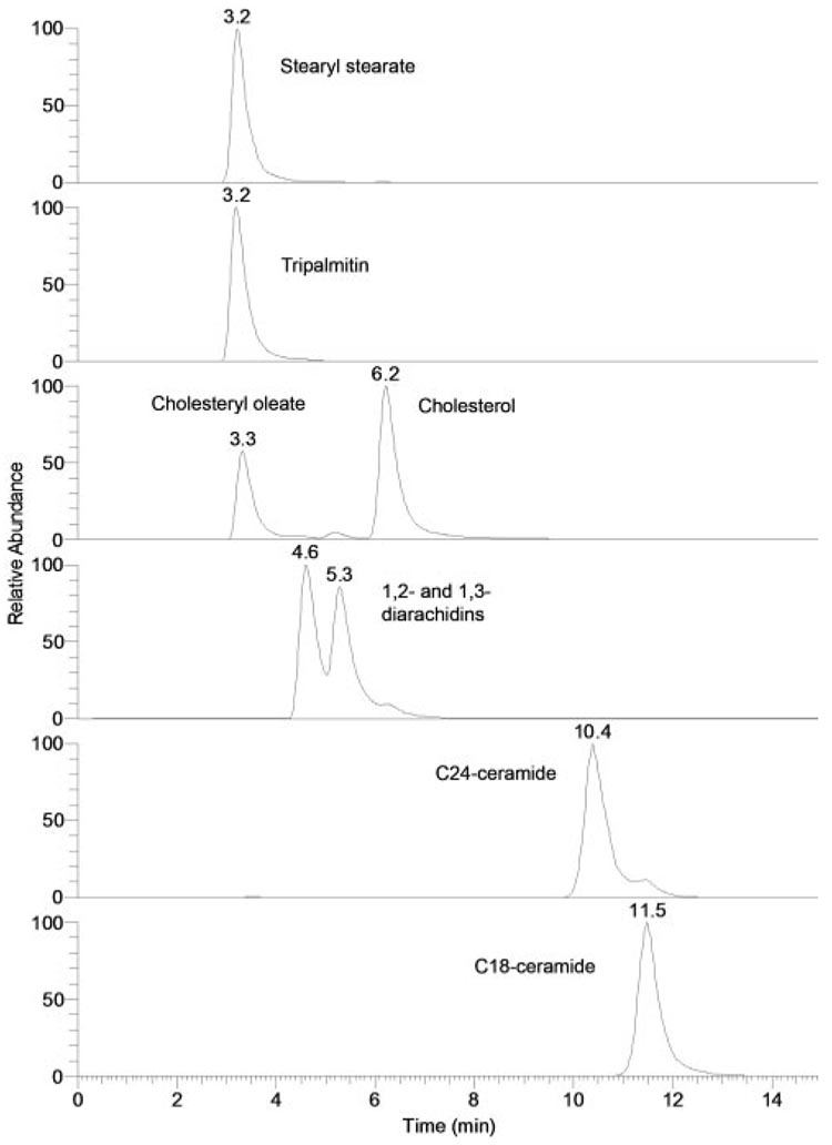
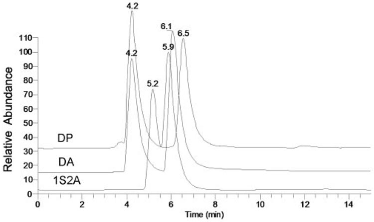
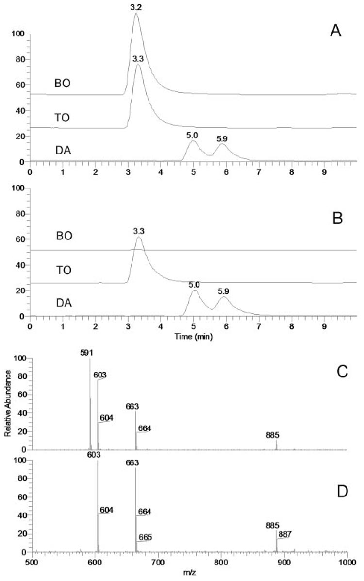
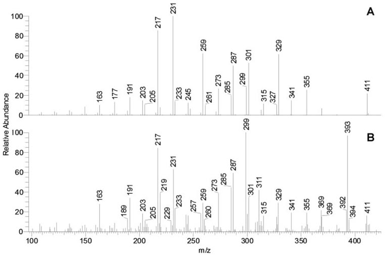
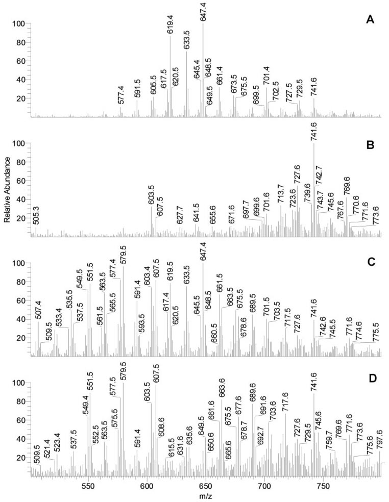
Methods: Samples T1 to T3 were analyzed with the use of high-pressure liquid chromatography/positive ion mode atmospheric pressure chemical ionization mass spectrometry. Where possible, the unknown lipids were compared with known standards.

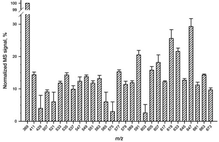
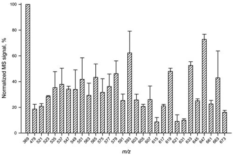
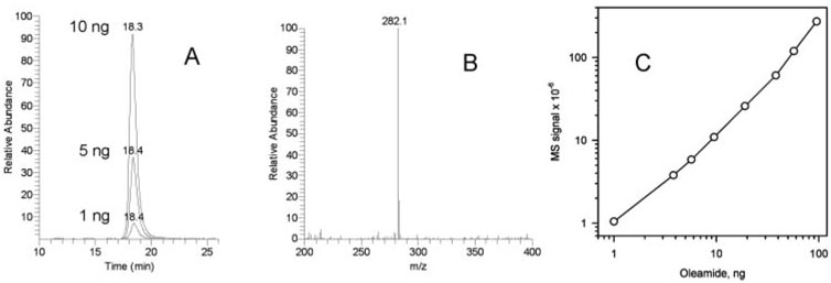
Results: Samples T1 had the simplest lipid composition among all the tested specimens. Samples T2 and T3 were similar to each other but were noticeably different from samples T1. In addition to all the compounds detected in samples T1, lower molecular weight wax esters and other compounds were found in samples T2 and T3. No appreciable amounts of fatty acid amides (e.g., oleamide), ceramides, or monoacyl glycerols were routinely detected. The occasionally observed minor signals of oleamide (m/z 282) in samples T3 were attributed to the contamination of the samples with common plasticizers routinely found in plastic ware extractives and organic solvents.

Conclusions: The MG is a prominent source of lipids for the tear film. However, it would have been a mistake to exclude from consideration other likely sources of lipids such as conjunctiva, cornea, and tears produced by the lacrimal glands. These data showed that lipids in AT are more complex than MG secretions, which necessitates more cautious interpretation of the functions of the latter in the tear film.


Figures

Figure 1
Figure 1
Human meibomian gland secretions (approximately 1 mg) at room temperature.
Figure 2
Figure 2
Melting curves of human meibomian gland secretions (A) and egg yolk phosphatidyl choline (B; mainly C16:0,C18:1 isomer).
Figure 3
Figure 3
Normal-phase HPLC and MS profiles of human meibomian gland secretions (T1) and aqueous tears (T2 and T3). (A) Total ion chromatogram of sample T1. (B) Total ion chromatogram of sample T2. (C) Total ion chromatogram of sample T3. (D) Combined MS profile of HPLC peaks with RT 3.6 and 4.3 minutes of sample T1. (E) Combined MS profile of HPLC peaks with RT 3.7 and 4.2 minutes of sample T2. (F) Combined MS profile of HPLC peaks with RT 3.6 and 4.2 minutes of sample T3.
Figure 4
Figure 4
Normal-phase HPLC-MS profiles of human aqueous (sample T3). (A) MS profile of HPLC peak with RT 3.6 minutes. (B) MS profile of HPLC peak with RT 4.2 minutes.
Figure 5
Figure 5
Reproducibility test for HPLC-APCI MS analysis of sample T2. Three sequential injections of the sample were made. Signals were normalized using the intensity of the cholesteryl signal (m/z 369) as 100%.
Figure 6
Figure 6
HPLC-APCI MS intersample variability test for sample T3. Seven individual samples T3 were analyzed. Signals were normalized using the intensity of the cholesteryl signal (m/z 369) in each of the samples as 100%.
Figure 7
Figure 7
Normal-phase HPLC elution profiles of nonpolar lipids. Chromatograms were plotted as single-ion chromatograms recorded in the positive ion mode: stearyl stearate (m/z 537; [M+H]+), tripalmitin (m/z 551, [M-palmitic acid+H]+), cholesteryl oleate and cholesterol (m/z 369; [M-oleic acid+H]+ and [M-H2O+H]+, respectively), 1,2- and 1,3-diarachidins (m/z 663; [M-H2O+H]+), C24-ceramide (m/z 632; [M-H2O+H]+), and C18-ceramide (m/z 548; [M-H2O+H]+).
Figure 8
Figure 8
Normal-phase HPLC elution profiles of standard diacyl glycerols dipalmitin (DP), diarachidin (DA), and 1-stearoyl-2-arachidonoyl glycerol (1S2A).
Figure 9
Figure 9
Effects of source fragmentation on HPLC elution profiles and mass spectra of wax esters and diacyl and triacyl glycerols. (A) Single-ion chromatograms of a mixture of behenyl oleate (BO; m/z 591; [M+H]+), trioleate (TO; m/z 603; [M-oleic acid+H]+), and diarachidin (DA; m/z 663; [M-H2O+H]+) without source fragmentation engaged. (B) Single-ion chromatograms of a mixture of BO, TO, and DA with source fragmentation (50 V) engaged. Note the absence of the HPLC peak of BO. (C) Integrated MS profile of HPLC peaks with RT 3.2 to 5.9 (no source fragmentation). (D) Integrated MS profile of HPLC peaks with RT 3.2 to 5.9 (source fragmentation at 50 V). Note the absence of the MS signal of BO (m/z 591).
Figure 10
Figure 10
Selective suppression of MS signals of wax esters in a mixture with diacyl and triacyl glycerols (5 µg/mL each) as a function of the fragmentation energy. (A) Absolute signal intensities of behenyl oleate (BO; m/z 591; [M+H]+), triolein (TO; m/z 885 and 603; [M+H]+ and [M-oleic acid+H]+, respectively), and diarachidin (DA; m/z 663; [M-H2O+H]+) at different fragmentation energy levels. (B) Relative intensities of the signals of BO, TO, and DA at different fragmentation energy levels. (C) Calculated ratios of the MS signals of BO, DA, and TO. Note that while the signal ratio DA/TO (663:885) remains fairly constant, the signal ratio BO/TO (591:603) becomes zero at the source fragmentation energy of 50 V, indicating complete suppression of the wax ester signal.
Figure 11
Figure 11
Mass spectra of typical impurities found in lipid samples. (A) Mass spectrum of contaminations in sample T3, which was stored in an Eppendorf tube. Signals 495, 551, 607, and 663 are typical of oxidized Irgafos 168, a common plasticizer used in the production of polyethylene. (B) Mass spectrum of an Irgafos 168 derivative (presumably a debutylated, methoxylated isomer; characteristic signals 581 and 637).
Figure 12
Figure 12
Detection and quantitation of oleamide. (A) Single-ion chromatogram of oleamide (m/z 282; [M+H]+). Injected amounts 1, 5, and 10 ng. (B) Mass spectrum of the HPLC peak of oleamide (RT, 18.3 minutes). (C) Calibration curve for oleamide quantitation. Note that no signals of oleamide were detected in most of the samples. One of five T1 samples showed oleamide in minor quantities (<0.05% of dry lipid weight). Two of thirteen T2 and T3 samples showed trace amounts of oleamide, but quantitation was not possible because of the uncertain dry lipid weight of samples T2 and T3.
Figure 13
Figure 13
Identification of squalene (m/z 411; [M+H]+) in aqueous tear samples (T3) in an MS/MS experiment. (A) Fragmentation of authentic squalene. (B) Fragmentation of ion m/z 411 detected in sample T3. Ion 393 is a signal of an unknown isobaric compound in the lipid mixture. Positiveion mode; solvent HPA; MS/MS fragmentation at relative energy of 38 V.
Figure 14
Figure 14
Selective suppression of wax ester signals in samples T1 and T3 to differentiate those from other components of lipid mixtures. (A) Mass spectrum of the nonpolar (WE, Chl-E, TAG) fraction of sample T1 recorded in an HPLC experiment (no source fragmentation engaged). (B) Mass spectrum of the nonpolar fraction of sample T1 recorded in an HPLC experiment (source fragmentation energy, 50 V). (C) Mass spectrum of the nonpolar fraction of sample T3 recorded in an HPLC experiment (no source fragmentation engaged). (D) Mass spectrum of the nonpolar fraction of sample T3 recorded in an HPLC experiment (source fragmentation energy, 50 V). Note that the WE signals (577–675) visible in MGs (A) were suppressed by engaging source fragmentation (B). A similar observation was made for WE in sample T3 (C, D).

References

    1. Andrews JS. Human tear film lipids, I: composition of the principal non-polar component. Exp Eye Res. 1970;10:223–227. - PubMed
    1. Brauninger GE, Shah DO, Kaufman HE. Direct physical demonstration of oily layer on tear film surface. Am J Ophthalmol. 1972;73:132–134. - PubMed
    1. McCulley JP, Shine W. A compositional based model for the tear film lipid layer. Trans Am Ophthalmol Soc. 1997;95:79–88. discussion 88–93. - PMC - PubMed
    1. Bron AJ, Tiffany JM, Gouveia SM, Yokoi N, Voon LW. Functional aspects of the tear film lipid layer. Exp Eye Res. 2004;78:347–360. - PubMed
    1. Shine WE, McCulley JP. Role of wax ester fatty alcohols in chronic blepharitis. Invest Ophthalmol Vis Sci. 1993;34:3515–3521. - PubMed

Publication types