. 2008 Jul;74(13):4059-69.
doi: 10.1128/AEM.00190-08. Epub 2008 May 16.

Identification of a cellobiose utilization gene cluster with cryptic beta-galactosidase activity in Vibrio fischeri

Affiliations

Identification of a cellobiose utilization gene cluster with cryptic beta-galactosidase activity in Vibrio fischeri

Dawn M Adin et al. Appl Environ Microbiol. 2008 Jul.

Abstract

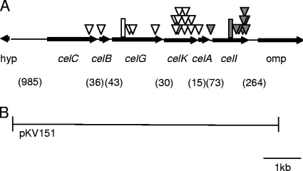
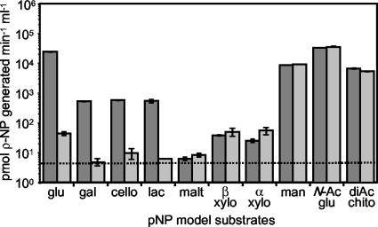
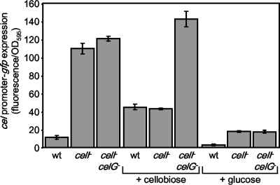
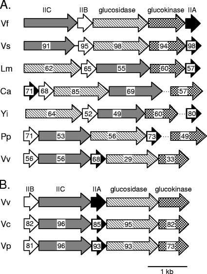
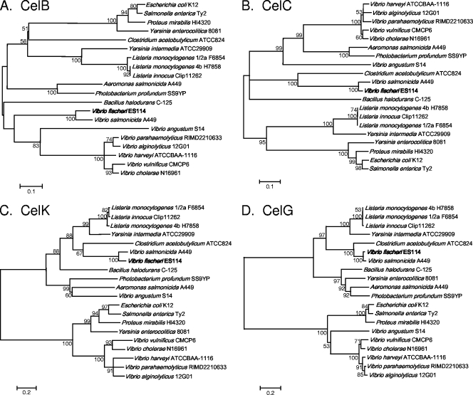
Cellobiose utilization is a variable trait that is often used to differentiate members of the family Vibrionaceae. We investigated how Vibrio fischeri ES114 utilizes cellobiose and found a cluster of genes required for growth on this beta-1,4-linked glucose disaccharide. This cluster includes genes annotated as a phosphotransferase system II (celA, celB, and celC), a glucokinase (celK), and a glucosidase (celG). Directly downstream of celCBGKA is celI, which encodes a LacI family regulator that represses cel transcription in the absence of cellobiose. When the celCBGKAI gene cluster was transferred to cellobiose-negative strains of Vibrio and Photobacterium, the cluster conferred the ability to utilize cellobiose. Genomic analyses of naturally cellobiose-positive Vibrio species revealed that V. salmonicida has a homolog of the celCBGKAI cluster, but V. vulnificus does not. Moreover, bioinformatic analyses revealed that CelG and CelK share the greatest homology with glucosidases and glucokinases in the phylum Firmicutes. These observations suggest that distinct genes for cellobiose utilization have been acquired by different lineages within the family Vibrionaceae. In addition, the loss of the celI regulator, but not the structural genes, attenuated the ability of V. fischeri to compete for colonization of its natural host, Euprymna scolopes, suggesting that repression of the cel gene cluster is important in this symbiosis. Finally, we show that the V. fischeri cellobioase (CelG) preferentially cleaves beta-d-glucose linkages but also cleaves beta-d-galactose-linked substrates such as 5-bromo-4-chloro-3-indolyl-beta-d-galactoside (X-gal), a finding that has important implications for the use of lacZ as a marker or reporter gene in V. fischeri.


Figures

FIG. 1.
FIG. 1.
Schematic representation of the genetic organization around the cel gene cluster in V. fischeri ES114. (A) Arrows represent ORFs and indicate the direction of gene transcription, as well as the gene size, which is presented relative to the scale bar. Shaded triangles represent transposon insertions that resulted in a blue colony phenotype on LBS-X-gal plates. Open triangles represent transposon insertions that resulted in a yellowish-white colony phenotype on LBS-X-gal cellobiose plates. A hypothetical gene is designated “hyp.” The shaded rectangle denotes the 11-bp deletion detected in mutants DMA428 and DMA429. The open rectangle denotes the 2-bp insertion in strain KV1319. Numbers in parentheses represent the base pairs between ORFs. (B) The region cloned into plasmid pKV151.
FIG. 2.
FIG. 2.
celG-dependent enzymatic activity. Activities in lysates from DMA420 (celI::mini-Tn5-Em) are shown as dark gray bars, and activities from DMA401 lysates (celG frameshift, celI::mini-Tn5-Em) are shown as light gray bars. Activities represent the ability to cleave pNP from the substrate. Error bars, some too small to visualize, indicate standard error (n = 3). The dashed line represents the limit of detection. Results shown are representative of two experiments at 37°C and are also similar to data obtained from two experiments at 28°C. Substrate abbreviations: glu, pNP-β-d-glucopyranoside (model substrate for cellobiose); gal, pNP-β-d-galactopyranoside; cello, pNP-β-d-cellobioside; lac, pNP-β-d-lactopyranoside; malt, pNP-β-d-maltoside; β xylo, pNP-β-d-xylopyranoside; α xylo, pNP-α-d-xylopyranoside; man, pNP-β-d-mannopyranoside; N-Ac glu, pNP-N-acetyl-β-d-glucosaminide; and diAC chito, pNP-N,N′-diacetyl-β-d-chitobioside.
FIG. 3.
FIG. 3.
Specific fluorescence generated from the Pcel-gfp reporter in ES114 (wild type [wt]), the mutant DMA420 (celI mutant), or DMA401 (celI celG double mutant). Cultures of strains carrying pDMA193 were grown in LBS with and without 10 mM cellobiose or 20 mM glucose. Data represent the average specific fluorescence with standard error (n = 3).
FIG. 4.
FIG. 4.
Homologs of the V. fischeri cellobiose-utilizing genes. Gene arrangement and orientation for each particular strain are indicated with arrows. Arrows with the same shading or pattern are putative homologs. Genes encoding PTS components A, B, and C are shaded black, white, or gray, respectively. Genes encoding glucokinases or glucosidases are filled with checkerboard or crosshatch patterns, respectively. Dotted lines indicate that glucokinase or PTS IIA genes in these bacteria are not genetically linked to the other genes shown. Numbers within the arrows represent the overall similarity to the respective protein from V. fischeri (panel A) or V. vulnificus (panel B) as determined by MatGAT software (10). Species abbreviations are: Vf, Vibrio fischeri; Vs, Vibrio salmonicida; Lm, Listeria monocytogenes; Ca, Clostridium acetobutylicum; Yi, Yersinia intermedia; Pp, Photobacterium profundum; Vv, Vibrio vulnificus; Vc, Vibrio cholerae; Vp, Vibrio parahaemolyticus.
FIG. 5.
FIG. 5.
Comparison of the V. fischeri CelB, CelC, CelK, and CelG proteins to proteins encoded by other bacteria with similar gene clusters. Consensus neighbor-joining trees constructed using MEGA 4.0 software (60) are shown. Trees constructed using both UPGMA and minimum evolution algorithms were similar to those shown in the figure (data not shown). Bootstrap values of >50% are indicated at the respective nodes. The scale bar represents a corrected sequence divergence of 0.1 or 0.2 as indicated. Trees in panels A, B, C, and D include V. fischeri proteins CelB, CelC, CelK, and CelG, respectively.

References

    1. Adin, D. M., N. J. Phillips, B. W. Gibson, M. A. Apicella, E. G. Ruby, M. J. McFall-Ngai, D. B. Hall, and E. V. Stabb. 2008. Characterization of htrB and msbB mutants of the light organ symbiont Vibrio fischeri. Appl. Environ. Microbiol. 74:633-644. - PMC - PubMed
    1. Aeckersberg, F., C. Lupp, B. Feliciano, and E. G. Ruby. 2001. Vibrio fischeri outer membrane protein OmpU plays a role in normal symbiotic colonization. J. Bacteriol. 183:6590-6597. - PMC - PubMed
    1. Altschul, S. F., W. Gish, W. Miller, E. W. Myers, and D. J. Lipman. 1990. Basic local alignment search tool. J. Mol. Biol. 215:403-410. - PubMed
    1. Antunes, L. C., A. L. Schaefer, R. B. Ferreira, N. Qin, A. M. Stevens, E. G. Ruby, and E. P. Greenberg. 2007. Transcriptome analysis of the Vibrio fischeri LuxR-LuxI regulon. J. Bacteriol. 189:8387-8391. - PMC - PubMed
    1. Ast, J. C., I. Cleenwerck, K. Engelbeen, H. Urbanczyk, F. L. Thompson, P. De Vos, and P. V. Dunlap. 2007. Photobacterium kishitanii sp. nov., a luminous marine bacterium symbiotic with deep-sea fishes. Int. J. Syst. Evol. Microbiol. 57:2073-2078. - PubMed

Publication types

LinkOut - more resources