. 2008 Jul;295(1):H227-36.
doi: 10.1152/ajpheart.01157.2007. Epub 2008 May 16.

Inhibition of brain proinflammatory cytokine synthesis reduces hypothalamic excitation in rats with ischemia-induced heart failure

Affiliations

Inhibition of brain proinflammatory cytokine synthesis reduces hypothalamic excitation in rats with ischemia-induced heart failure

Yu-Ming Kang et al. Am J Physiol Heart Circ Physiol. 2008 Jul.

Abstract

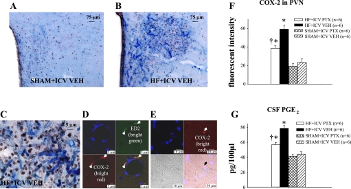
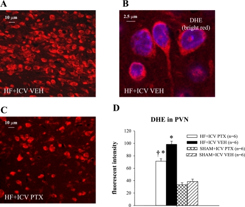
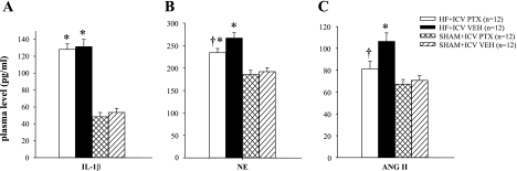
The expression of proinflammatory cytokines increases in the hypothalamus of rats with heart failure (HF). The pathophysiological significance of this observation is unknown. We hypothesized that hypothalamic proinflammatory cytokines upregulate the activity of central neural systems that contribute to increased sympathetic nerve activity in HF, specifically, the brain renin-angiotensin system (RAS) and the hypothalamic-pituitary-adrenal (HPA) axis. Rats with HF induced by coronary ligation and sham-operated controls (SHAM) were treated for 4 wk with a continuous intracerebroventricular infusion of the cytokine synthesis inhibitor pentoxifylline (PTX, 10 microg/h) or artificial cerebrospinal fluid (VEH). In VEH-treated HF rats, compared with VEH-treated SHAM rats, the hypothalamic expression of proinflammatory cytokines was increased, along with key components of the brain RAS (renin, angiotensin-converting enzyme, angiotensin type 1 receptor) and corticotropin-releasing hormone, the central indicator of HPA axis activation, in the paraventricular nucleus (PVN) of the hypothalamus. The expression of other inflammatory/excitatory mediators (superoxide, prostaglandin E(2)) was also increased, along with evidence of chronic neuronal excitation in PVN. VEH-treated HF rats had higher plasma levels of norepinephrine, ANG II, interleukin (IL)-1beta, and adrenocorticotropic hormone, increased left ventricular end-diastolic pressure, and increased wet lung-to-body weight ratio. With the exception of plasma IL-1beta, an indicator of peripheral proinflammatory cytokine activity, all measures of neurohumoral excitation were significantly lower in HF rats treated with intracerebroventricular PTX. These findings suggest that the increase in brain proinflammatory cytokines observed in rats with ischemia-induced HF is functionally significant, contributing to neurohumoral excitation by activating brain RAS and the HPA axis.


Figures

Fig. 1.
Fig. 1.
Group data showing the effect of treatment with intracerebroventricular (ICV) PTX on hypothalamic tissue levels of proinflammatory cytokines and on neuronal activity in paraventricular nucleus (PVN) of hypothalamus in rats with ischemia-induced heart failure (HF). A: tumor necrosis factor-α (TNF-α) and interleukin-1β (IL-1β) increased in hypothalamus of vehicle (VEH)-treated HF rats compared with VEH-treated sham-operated (SHAM) rats. TNF-α and IL-1β levels were less in pentoxifylline (PTX)-treated than VEH-treated HF rats. B: Fra-LI activity, an indicator of chronic neuronal excitation, increased in the PVN of VEH-treated HF rats compared with VEH-treated SHAM. Fra-LI activity was less in the PTX-treated than VEH-treated HF rats. P < 0.05 vs. SHAM + VEH (*) and HF + PTX vs. HF + VEH (†).
Fig. 2.
Fig. 2.
Expression of cyclooxygenase-2 (COX-2) and prostaglandin E2 (PGE2) in the brain. Double-labeling immunohistochemistry showing COX-2 (blue) and Fra-LI activity (black dots), an indicator of chronic neuronal excitation, in a coronal section of the PVN of a SHAM rat (A) and a HF rat (B). C: high-power view of the section shown in B, demonstrating COX-2 activity in the microvasculature penetrating the PVN, closely associated with Fra-LI positive PVN neurons. D: confocal images demonstrating triple immunostaining for nuclei (blue), the perivascular cell marker ED2 (bright green), and COX-2 (bright red) in the PVN. The yellow color in the combined image confirms dual labeling for ED2 and COX-2. E: confocal images showing endothelial cells (blue) and COX-2 (bright red), with an apparent localization of COX-2 in perivascular rather than endothelial cells in the microvasculature of PVN. F: group data showing effect of ICV PTX on COX-2 fluorescent intensity in PVN of HF and SHAM rats. COX-2 immunofluorescence was reduced in PTX-treated HF rats. G: group data showing cerebrospinal fluid (CSF) levels of PGE2, a physiological indicator of COX-2 activity, increase in VEH-treated HF rats compared with VEH-treated SHAM. The CSF PGE2 level is lower in the PTX-treated rats. P < 0.05 vs. SHAM + VEH (*) and HF + PTX vs. HF + VEH (†).
Fig. 3.
Fig. 3.
Immunohistochemistry for corticotropin-releasing hormone (CRH) expression in the PVN of hypothalamus. A: double labeling for CRH (red) and Fra-LI activity (black dots), an indicator of chronic neuronal excitation, in a coronal section of the PVN of a HF rat. B: high-power view of the section shown in A demonstrating CRH labeling of the cytoplasm of a Fra-LI positive PVN neuron. C: effect of ICV PTX treatment on CRH expression and Fra-LI activity in PVN of a HF rat. D: group data showing effects of ICV PTX on numbers of PVN neurons positive for Fra-LI activity that also express CRH in HF and SHAM rats. E: group data showing effects of ICV PTX on plasma levels of ACTH in HF and SHAM rats. P < 0.05 vs. SHAM + VEH (*) and HF + PTX vs. HF + VEH (†).
Fig. 4.
Fig. 4.
Immunofluorescence for renin and angiotensin-converting enzyme (ACE) in the parvocellular region of the PVN of hypothalamus. A: coronal section of the PVN of a VEH-treated HF rat showing immunofluorescence for renin (red) and neuronal nuclei (blue). Side panel shows magnified image. B: coronal section of the PVN of a PTX-treated HF rat showing reduced immunofluorescence for renin. C: group data showing effect of ICV PTX on fluorescent intensity of renin in PVN of HF and SHAM rats. D: coronal section of the PVN of a VEH-treated HF rat showing immunofluorescence for ACE (red) and neuronal nuclei (blue). Side panel shows magnified image. E: coronal section of the PVN of a PTX-treated HF rat showing reduced immunofluorescence for ACE. F: group data showing effect of ICV PTX on fluorescent intensity of ACE in PVN of HF and SHAM rats. P < 0.05 vs. SHAM + VEH (*) and HF + PTX vs. HF + VEH (†).
Fig. 5.
Fig. 5.
Immunohistochemistry for ACE expression in the PVN of hypothalamus. A: immunohistochemical double-labeling for ACE (red) and Fra-LI activity (black dots), an indicator of chronic neuronal excitation, in a coronal section of the PVN of a HF rat. B: high-power image taken from the section shown in A demonstrating ACE labeling of the cytoplasm of a Fra-LI-positive PVN neuron. C: effect of ICV PTX treatment on ACE expression in Fra-LI-positive PVN neurons of a HF rat. D: group data showing effects of ICV PTX on numbers of Fra-LI-positive neurons also expressing ACE in PVN of HF and SHAM rats. P < 0.05 vs. SHAM + VEH (*) and HF + PTX vs. HF + VEH (†).
Fig. 6.
Fig. 6.
Angiotensin type 1 receptor (AT1-R) expression in the parvocellular region of the PVN of hypothalamus. A: immunofluorescence for AT1-R (red) and neuronal nuclei (blue) in a coronal section of the PVN of a VEH-treated HF rat. Side panel shows magnified image. B: immunofluorescence for AT1-R (red) and neuronal nuclei (blue) in a coronal section of the PVN of a PTX-treated HF rat. C: group data showing effect of ICV PTX on fluorescent intensity of AT1-R in PVN of HF and SHAM rats. D: group data showing AT1-R protein in the hypothalamus of ICV PTX- and VEH-treated HF and SHAM rats. Representative Western blots are shown above the bar graph. P < 0.05 vs. SHAM + VEH (*) and HF + PTX vs. HF + VEH (†).
Fig. 7.
Fig. 7.
Superoxide expression in the parvocellular region of the PVN of hypothalamus. A: immunofluorescence for superoxide in PVN of a HF rat indicated by dihydroethidium (DHE) staining (red). B: high-power image taken from the section shown in A. C: DHE labeling in the PVN of a PTX-treated HF rat. D: group data showing effect of ICV PTX on fluorescent intensity of DHE in PVN of HF and SHAM rats. P < 0.05 vs. SHAM + VEH (*) and HF + PTX vs. HF + VEH (†).
Fig. 8.
Fig. 8.
Humoral indicators of HF. Plasma levels of IL-1β (A), norepinephrine (NE; B) and ANG II (C) in HF and SHAM rats treated for 4 wk with ICV PTX or VEH. NE and ANG II were lower in HF rats treated with ICV PTX, but IL-1β was unaffected. P < 0.05 vs. SHAM + VEH (*) and HF + PTX vs. HF + VEH (†).

Similar articles

Cited by

References

    1. American Physiological Society. Guiding principles for research involving animals and human beings. Am J Physiol Regul Integr Comp Physiol 283: R281–R283, 2002. - PubMed
    1. Bienvenu J, Doche C, Gutowski MC, Lenoble M, Lepape A, Perdrix JP. Production of proinflammatory cytokines and cytokines involved in the TH1/TH2 balance is modulated by pentoxifylline. J Cardiovasc Pharmacol 25, Suppl 2: S80–S84, 1995. - PubMed
    1. Bristow MR Why does the myocardium fail? Insights from basic science. Lancet 352, Suppl 1: SI8–SI14, 1998. - PubMed
    1. Brown MR, Fisher LA. Corticotropin-releasing factor: effects on the autonomic nervous system and visceral systems. Fed Proc 44: 243–248, 1985. - PubMed
    1. Catt KJ, Millan MA, Wynn PC, Mendelsohn FA, Aguilera G. Brain receptors for hypothalamic hormones. Adv Biochem Psychopharmacol 43: 51–67, 1987. - PubMed

Publication types

MeSH terms