. 2008 Sep;7(9):1587-97.
doi: 10.1074/mcp.M800113-MCP200. Epub 2008 May 16.

Prevention of amino acid conversion in SILAC experiments with embryonic stem cells

Affiliations

Prevention of amino acid conversion in SILAC experiments with embryonic stem cells

Sean C Bendall et al. Mol Cell Proteomics. 2008 Sep.

Abstract

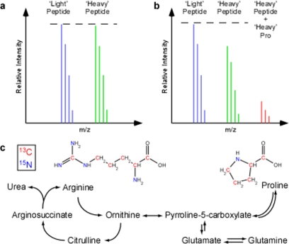
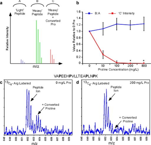
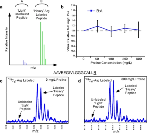
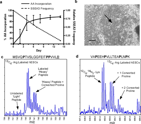
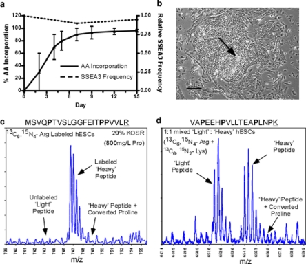
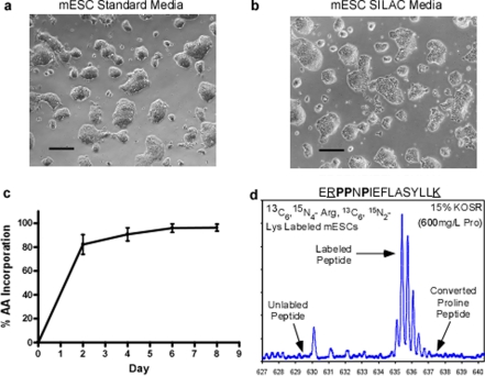
Recent studies using stable isotope labeling with amino acids in culture (SILAC) in quantitative proteomics have made mention of the problematic conversion of isotope-coded arginine to proline in cells. The resulting converted proline peptide divides the heavy peptide ion signal causing inaccuracy when compared with the light peptide ion signal. This is of particular concern as it can effect up to half of all peptides in a proteomic experiment. Strategies to both compensate for and limit the inadvertent conversion have been demonstrated, but none have been shown to prevent it. Additionally, these methods combined with SILAC labeling in general have proven problematic in their large scale application to sensitive cell types including embryonic stem cells (ESCs) from the mouse and human. Here, we show that by providing as little as 200 mg/liter L-proline in SILAC media, the conversion of arginine to proline can be rendered completely undetectable. At the same time, there was no compromise in labeling with isotope-coded arginine, indicating there is no observable back conversion from the proline supplement. As a result, when supplemented with proline, correct interpretation of "light" and "heavy" peptide ratios could be achieved even in the worst cases of conversion. By extending these principles to ESC culture protocols and reagents we were able to routinely SILAC label both mouse and human ESCs in the absence of feeder cells and without compromising the pluripotent phenotype. This study provides the simplest protocol to prevent proline artifacts in SILAC labeling experiments with arginine. Moreover, it presents a robust, feeder cell-free, protocol for performing SILAC experiments on ESCs from both the mouse and the human.


Figures

F<sc>ig</sc>. 1.
Fig. 1.
Metabolic conversion of isotope-coded arginine to proline in SILAC experiments. The unpredictable conversion of isotopic arginine to proline creates inaccuracy in SILAC-based quantitative proteomic experiments. a, a conceptual mass spectra of a non-proline containing peptide ion from a 1:1 mixture of light and heavy labeled samples. Here the expected Light and Heavy peptide ions have an equivalent signal. b, a spectra from a proline containing peptide where arginine to proline conversion has occurred in the same 1:1 mixture. The resulting heavy proline peptide signal (red) has been subtracted from the expected ‘heavy’ peptide ion signal. c, metabolic pathway outlining the inter-conversion of arginine and proline. Isotope-coded arginine with carbon 13 (red) and nitrogen 15 (green) when used as a synthetic precursor increases the expected mass of proline.
F<sc>ig</sc>. 2.
Fig. 2.
Titration of proline during SILAC labeling with isotope-coded arginine. Candidate proline containing tryptic peptides were monitored in whole cell extracts in 13C6-arginine samples with 0–800 mg/liter l-proline. a and b, conceptual mass spectra indicating the possible peptide ion peaks observed in the arginine labeling experiment. Both the ratio of labeled to unlabeled peptide as well as relative intensity of converted proline peptide ion was monitored in the experiment across 0–800 mg/liter proline (b) where * indicates no converted proline peptide ions were detectable from background. c, a mass spectrum from 0 mg/liter proline sample representing the peptide VAPEEHPVLLTEAPINPK at m/z 652 demonstrating a prevalent converted proline peak at +1.7 m/z from the expected peptide ion. d, mass spectrum from the 200 mg/liter proline sample depicting the same peptide ion as shown in c and the converted proline ion is notably absent.
F<sc>ig</sc>. 3.
Fig. 3.
Isotope-coded arginine labeling efficiency across the titration of proline. Candidate arginine containing tryptic peptides lacking proline was monitored in whole cell extracts in 13C6-arginine samples with 0–800 mg/liter l-proline. a, conceptual mass spectra indicating the possible peptide ion peaks observed in the arginine labeling experiment. The peak area ratio between the heavy and light arginine monoisotopic peaks was monitored across the 0–800 mg/liter proline samples and plotted relative to 0 mg/liter proline (b). c, a mass spectrum representing the peptide AAVEEGIVLGGGCALLR at m/z 845 from 0 mg/liter proline sample. d, mass spectra of the same peptide ion in the 800 mg/liter proline sample demonstrating no compromise in the isotope-coded arginine labeling efficiency or proline to arginine back conversion. Isotopic amino acids are underlined.
F<sc>ig</sc>. 4.
Fig. 4.
Labeling feeder cell-free hESCs in standard SILAC medium. hESCs culture on Matrigel was grown in standard SILAC medium (13C6-Arg, dialyzed FBS, 0 mg/liter proline) for 2 weeks. Arginine incorporation, conversion to proline, and hESC phenotype were monitored. a, the relative SSEA3 frequency and percent heavy arginine incorporation over the SILAC labeling time course. b, differentiated phenotype of hESC after 14 days in standard SILAC medium as observed by light microscopy. Scale bars equal 250 μm. c, a mass spectrum of heavy arginine peptide ion MSVQPTVSLGGFEITPPVLR at 745 m/z. Indicated are the prominent converted proline peptide ion shifted +1.7 m/z and the absence of the unlabeled light arginine peptide ion corresponding to the high level of isotopic amino acid incorporation. d, a mass spectrum of the peptide ion VAPEEHPVLLTEAPLNPK at 652 m/z. Indicated are the peptide ions shifted +1.7 m/z and +3.4 m/z corresponding to the incorporation of 1 and 2 converted heavy proline and consuming more than half of the expected peptide ion intensity. Isotopic amino acids are underlined.
F<sc>ig</sc>. 5.
Fig. 5.
Labeling feeder cell-free hESCs in conditioned SILAC medium with KOSR. hESCs cultured on Matrigel were grown in MEF-conditioned ESC optimized SILAC media (13C6 15N4-Arg and 13C6, 15N2-Lys, 20% KOSR providing 800 mg/liter l-proline) for 2 weeks. Amino acid incorporation, conversion of arginine to proline, and hESC phenotype were monitored. a, the relative SSEA3 frequency and percent heavy amino acid incorporation over the SILAC labeling time course. b, undifferentiated phenotype maintained by hESCs after more than 15 days in optimized SILAC media as observed by light microscopy. Scale bars equal 250 μm. c, a mass spectrum of heavy arginine peptide ion MSVQPTVSLGGFEITPPVLR at ∼747 m/z. Indicated are the absence of both a converted proline peptide ion and the unlabeled light arginine peptide ion. The absent light peptide ion at 743 m/z also indicates no back conversion of proline to unlabeled arginine. d, a mass spectrum of the 1:1 mixed labeled and unlabeled hESC lysates showing light and heavy peptide ion VAPEEHPVLLTEAPLNPK at ∼652 m/z and 655 m/z. The prominent converted proline peptide ions are now almost undetectable and do not effect the observed 1:1 ratio. Isotopic amino acids are underlined.
F<sc>ig</sc>. 6.
Fig. 6.
Labeling feeder cell-free mESCs in conditioned SILAC media with KOSR. mESCs cultured on gelatin were grown in ESC-optimized SILAC media (13C6, 15N4-Arg and 13C6, 15N2-Lys, 15% KOSR providing 600 mg/liter l-proline) for 4 passages (8 days). Amino acid incorporation, conversion of arginine to proline, and mESC phenotype were monitored. The undifferentiated phenotype of mESCs grown under standard feeder cell-free conditions (a) compared with those grown in optimized SILAC medium for 8 days (b) as observed by light microscopy. Scale bars equal 250 μm. c, the average heavy Lys and Arg incorporation over the SILAC labeling time course. d, a mass spectrum of peptide ERPPNPIEFLASYLLK at 635.6 m/z from mESCs in ESC-optimized SILAC media. The heavy Lys and Arg peptide ions are prominent whereas the unlabeled light peptide or converted heavy proline peptide ions are absent. The absent light peptide ion at m/z 633 also indicates no back conversion of proline to unlabeled arginine. Isotopic amino acids are underlined.

References

    1. Aebersold, R., and Mann, M. ( 2003) Mass spectrometry-based proteomics. Nature 422, 198–207 - PubMed
    1. Ong, S. E., and Mann, M. ( 2005) Mass spectrometry-based proteomics turns quantitative. Nat. Chem. Biol. 1, 252–262 - PubMed
    1. Hwang, S. I., Lundgren, D. H., Mayya, V., Rezaul, K., Cowan, A. E., Eng, J. K., and Han, D. K. ( 2006) Systematic characterization of nuclear proteome during apoptosis: a quantitative proteomic study by differential extraction and stable isotope labeling. Mol. Cell Proteomics 5, 1131–1145 - PubMed
    1. Ong, S. E., Kratchmarova, I., and Mann, M. ( 2003) Properties of 13C-substituted arginine in stable isotope labeling by amino acids in cell culture (SILAC). J. Proteome Res. 2, 173–181 - PubMed
    1. Ong, S. E., and Mann, M. ( 2006) A practical recipe for stable isotope labeling by amino acids in cell culture (SILAC). Nat. Protoc. 1, 2650–2660 - PubMed

Publication types