. 2008 Aug;7(8):1541-55.
doi: 10.1074/mcp.M800007-MCP200. Epub 2008 May 16.

Targets of caspase-6 activity in human neurons and Alzheimer disease

Affiliations

Targets of caspase-6 activity in human neurons and Alzheimer disease

Guy Klaiman et al. Mol Cell Proteomics. 2008 Aug.

Abstract

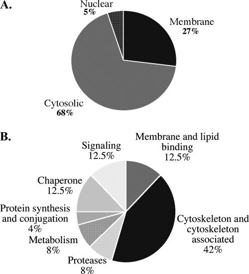
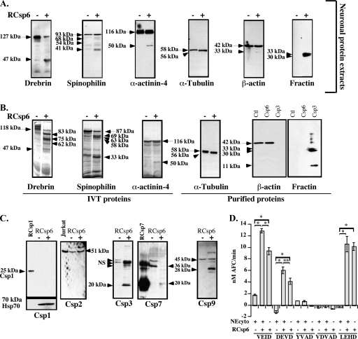
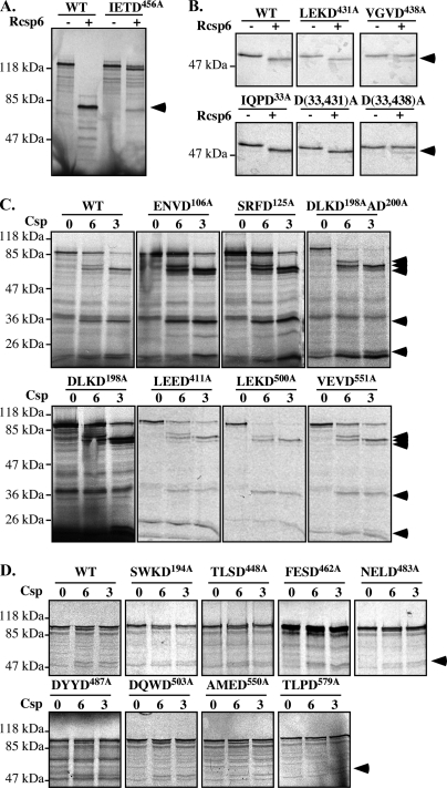
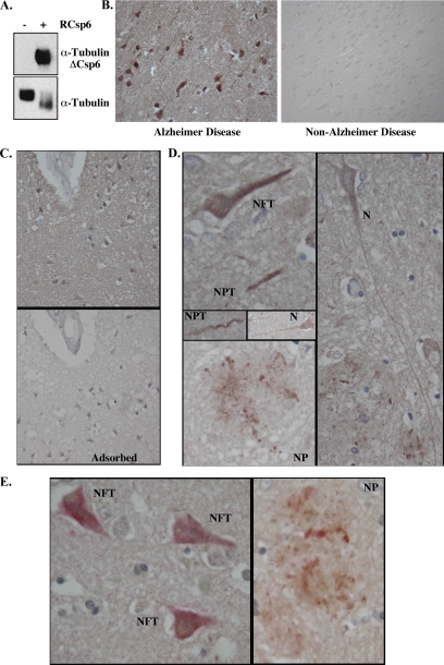
Caspase-6 activation occurs early in Alzheimer disease and sometimes precedes the clinical manifestation of the disease in aged individuals. The active Caspase-6 is localized in neuritic plaques, in neuropil threads, and in neurofibrillary tangles containing neurons that are not morphologically apoptotic in nature. To investigate the potential consequences of the activation of Caspase-6 in neurons, we conducted a proteomics analysis of Caspase-6-mediated cleavage of human neuronal proteins. Proteins from the cytosolic and membrane subcellular compartments were treated with recombinant active Caspase-6 and compared with undigested proteins by two-dimensional gel electrophoresis. LC/MS/MS analyses of the proteins that were cleaved identified 24 different potential protein substrates. Of these, 40% were cytoskeleton or cytoskeleton-associated proteins. We focused on the cytoskeleton proteins because these are critical for neuronal structure and function. Caspase-6 cleavage of alpha-Tubulin, alpha-Actinin-4, Spinophilin, and Drebrin was confirmed. At least one Caspase-6 cleavage site was identified for Drebrin, Spinophilin, and alpha-Tubulin. A neoepitope antiserum to alpha-Tubulin cleaved by Caspase-6 immunostained neurons, neurofibrillary tangles, neuropil threads, and neuritic plaques in Alzheimer disease and co-localized with active Caspase-6. These results imply that the early and neuritic activation of Caspase-6 in Alzheimer disease could disrupt the cytoskeleton network of neurons, resulting in impaired neuronal structure and function in the absence of cell death. This study provides novel insights into the pathophysiology of Alzheimer disease.


Figures

F<sc>ig</sc>. 1.
Fig. 1.
Purification of subcellular fractions from primary human neurons. A, micrograph of MAP2 immunofluorescent staining (upper panel) and micrograph overlay of MAP2 fluorescent staining and phase contrast (lower panel) of the human neuronal cultures. B, Western blot analysis of primary human neuron subcellular fractions probed with mouse anti-cytosolic Hsp70 (cHsp70), mouse anti-mitochondrial Hsp70 (mtHsp70), and rabbit anti-PARP antibodies.
F<sc>ig</sc>. 2.
Fig. 2.
Two-dimensional gel analysis of Csp6-cleaved proteins. Shown is a comparison of 2D gels: non-digested (no RCsp6) versus RCsp6 (+RCsp6)-digested proteins extracted from the cytosolic (A) and membrane (B) subcellular fractions. Spots that disappeared with RCsp6 are labeled with an arrow, spots that decreased with RCsp6 are labeled with a block arrow, and spots that increased with RCsp6 are indicated with a rounded arrow. C, high magnification of some of the spots that disappeared (encircled) shown in A.
F<sc>ig</sc>. 3.
Fig. 3.
Schematic diagram of proteomics analysis results. A, pie chart showing the percentage of proteins identified as putative Csp6 substrates in the membrane, cytosolic, and nuclear subcellular fractions of primary cultures of human neurons. B, pie chart of the various functional categories of putative Csp6 substrates. The percentage of proteins in each category is indicated.
F<sc>ig</sc>. 4.
Fig. 4.
Cleavage of specific proteins by Csp6. A and B, Western blot analyses or autoradiogram (for IVT Drebrin, Spinophilin, and α-Actinin-4) of neuronal protein extracts (A) or IVT or purified proteins (B) cleaved with RCsp6. The arrows indicate full-length proteins, and the arrowheads indicate the fragments. C, Western blot analysis of Csp1, Csp2, Csp3, Csp7, and Csp9 in 50 μg of neuronal cytosolic proteins treated without (−) or with (+) RCsp6. D, fluorogenic caspase activity assays of 1 μg of cytosolic neuronal protein extracts (NEcyto) treated with 2.7 ng of RCsp6 on YVAD-AFC for Csp1, VDVAD-AFC for Csp2, DEVD-AFC for Csp3 and Csp7, VEID-AFC for Csp6 and Csp8, and LEHD-AFC for Csp9. * indicates a p < 0.001 and ** indicates a p < 0.01 statistically significant difference between the indicated assays. NS, nonspecific. Error bars indicate standard deviation.
F<sc>ig</sc>. 5.
Fig. 5.
Identification of Csp6 cleavage sites. Shown are autoradiograms of IVT WT and mutant D456A Drebrin (A) and WT and mutant D431A, D438A, D33A, D33A/D431A (D(33,431)A), and D33A/D438A (D(33,438)A) α-Tubulin (B) before (−) and after (+) Csp6 cleavage. C and D, autoradiograms of IVT WT and mutant Spinophilin (C) and α-Actinin-4 (D) before (0) and after cleavage with Csp6 (6) and Csp3 (3). The arrowheads indicate cleaved fragments.
F<sc>ig</sc>. 6.
Fig. 6.
Identification α-Tubulin cleaved by Csp6 in AD brains. A, Western blot analysis of purified Tubulin without or with RCsp6 cleavage using Tubulin cleaved by Csp6 (anti-TubulinΔCsp6) and Tubulin antisera. B, micrographs of immunohistochemical detection of anti-TubulinΔCsp6 with diaminobenzidine in tissue sections of AD and non-AD temporal cortex. C, micrograph of immunohistochemical detection of anti-TubulinΔCsp6 in AD tissue sections showing the adsorption of the antiserum on the antigenic peptide. D, micrographs of α-TubulinΔCsp6 immunopositive NFT, neuropil threads (NPT), NP, and neurons (N) that otherwise look normal. E, micrograph of double immunostaining of TubulinΔCsp6 with diaminobenzidine (brown) and active Csp6 with fast red (pink) in NFT and NP.

References

    1. Stadelmann, C., Deckwerth, T., Srinivasan, A., Bancher, C., Bruck, W., Jellinger, K., and Lassmann, H. ( 1999) Activation of caspase-3 in single neurons and autophagic granules of granulovacuolar degeneration in Alzheimer's disease. Am. J. Pathol. 155, 1459–1466 - PMC - PubMed
    1. Selznick, L., Holtzman, D., Han, B., Gokden, M., Srinivasan, A., Johnson, E., and Roth, K. ( 1999) In situ immunodetection of neuronal caspase-3 activation in Alzheimer's disease. J. Neuropathol. Exp. Neurol. 58, 1020–1026 - PubMed
    1. Guo, H., Albrecht, S., Bourdeau, M., Petzke, T., Bergeron, C., and LeBlanc, A. C. ( 2004) Active Caspase-6 and Caspase-6 cleaved Tau in neuropil threads, neuritic plaques and neurofibrillary tangles of Alzheimer's disease. Am. J. Pathol. 165, 523–531 - PMC - PubMed
    1. Albrecht, S., Bourdeau, M., Bennett, D., Mufson, E. J., Bhattacharjee, M., and LeBlanc, A. C. ( 2007) Activation of caspase-6 in aging and mild cognitive impairment. Am. J. Pathol. 170, 1200–1209 - PMC - PubMed
    1. Ruchaud, S., Korfali, N., Villa, P., Kottke, T. J., Dingwall, C., Kaufmann, S. H., and Earnshaw, W. C. ( 2002) Caspase-6 gene disruption reveals a requirement for lamin A cleavage in apoptotic chromatin condensation. EMBO J. 21, 1967–1977 - PMC - PubMed

Publication types