Comparative Study
. 2008 Jun;9(2):133-44.
doi: 10.4142/jvs.2008.9.2.133.

The C-terminal variable domain of LigB from Leptospira mediates binding to fibronectin

Affiliations
Comparative Study

The C-terminal variable domain of LigB from Leptospira mediates binding to fibronectin

Yi-Pin Lin et al. J Vet Sci. 2008 Jun.

Abstract

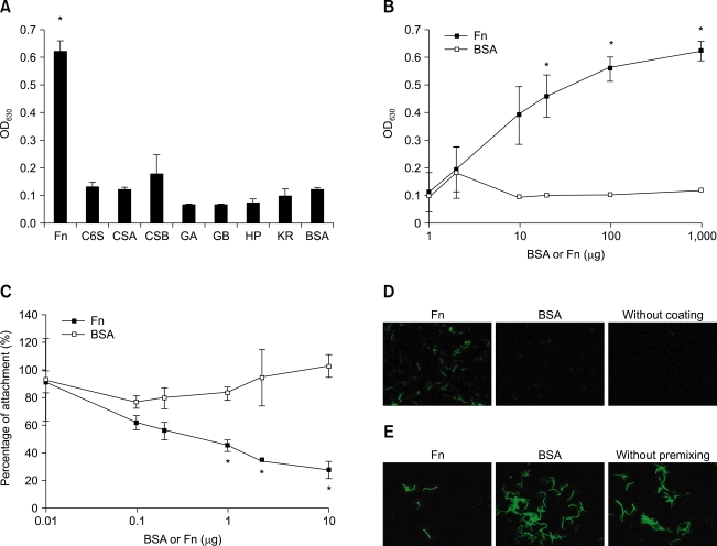
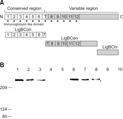
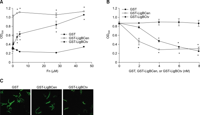
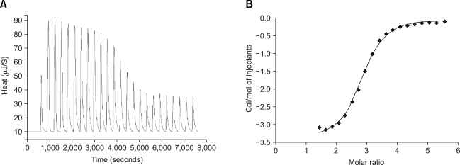
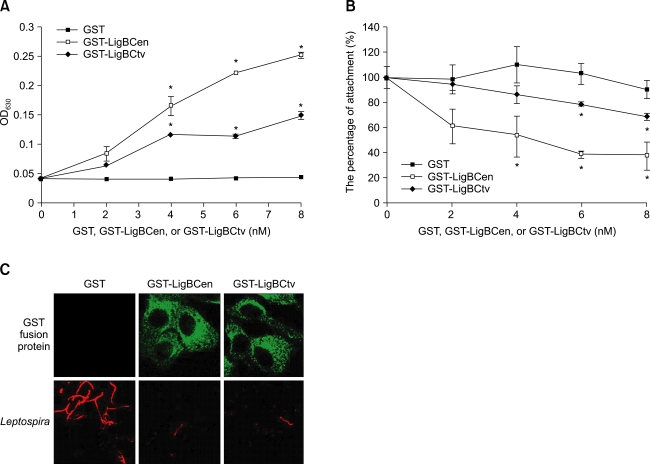
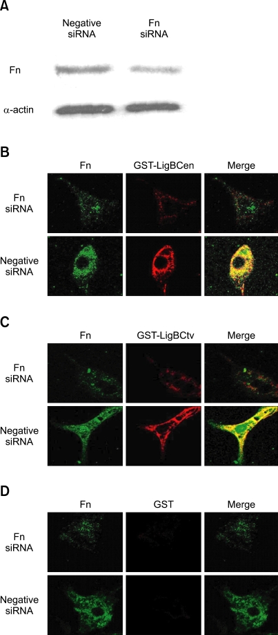
Adhesion through microbial surface components that recognize adhesive matrix molecules is an essential step in infection for most pathogenic bacteria. In this study, we report that LigB interacts with fibronectin (Fn) through its variable region. A possible role for LigB in bacterial attachment to host cells during the course of infection is supported by the following observations: (i) binding of the variable region of LigB to Madin-Darby canine kidney (MDCK) cells in a dose-dependent manner reduces the adhesion of Leptospira, (ii) inhibition of leptospiral attachment to Fn by the variable region of LigB, and (iii) decrease in binding of the variable region of LigB to the MDCK cells in the presence of Fn. Furthermore, we found a significant reduction in binding of the variable region of LigB to Fn using small interfering RNA (siRNA). Finally, the isothermal titration calorimetric results confirmed the interaction between the variable region of LigB and Fn. This is the first report to demonstrate that LigB binds to MDCK cells. In addition, the reduction of Fn expression in the MDCK cells, by siRNA, reduced the binding of LigB. Taken together, the data from the present study showed that LigB is a Fn-binding protein of pathogenic Leptospira spp. and may play a pivotal role in Leptospira-host interaction during the initial stage of infection.


Figures

Fig. 1
Fig. 1
The binding of L. interrogans serovar Pomona (NVSL 1427-35-093002) to Fn (A). Binding of Leptopsira to various immobilized ECM components. Leptospira (107) were added to wells coated with each ECM (1 mg in 100 µl PBS) including Fn, chondroitin-6-sulfate (C6S), chondroitin sulfate A (CSA), chondroitin sulfate B (CSB), gelatin A (GA), gelatin B (GB), heparin (HP), keratin (KR), or BSA (negative control). (B). Binding of Leptospira (107) to various concentrations of Fn (0, 10, 20, 100 or 1,000 µg in 100 µl PBS). BSA served as a negative control. (C). Fn inhibits the binding of Leptospira to the MDCK cells. Leptospira (107) were treated with various concentrations of Fn (0, 0.01, 0.1, 0.2, 1, 2, or 10 µg) or BSA (negative control) prior to addition to the MDCK cells (105). The percentage adhesion was determined relative to the attachment of untreated Leptospira onto the MDCK cells. (D). Binding of Leptospira to immobilize Fn. Leptospira (108) were cultured in Fn or BSA (negative control) coated (1 mg in 100 µl PBS) or un-coated wells (negative control). (E). Fn inhibited the binding of Leptospira to the MDCK cells. Leptospira (108) were pre-treated with 10 µg of Fn or BSA (negative control) prior to addition to the MDCK cells (106). Un-treated Leptospira was used as a negative control. The binding of Leptospira to ECMs or Fn or the adhesion of Leptospira to the MDCK cells was measured by ELISA (A, B, and C) or EPM (D and E). For all experiments, each value represents the mean ± SE of three trials performed in triplicate samples. Statistically significant (p < 0.05) differences are indicted by an asterisk. The EPM settings were identical for all captured images (D and E).
Fig. 2
Fig. 2
The interaction between LigB and Fn by the GST-pull down assay (A) A schematic diagram showing the structure of LigB and the truncated LigB protein used in this study. (B). Human plasma Fn ( lane 2 to lane 5 ) or cell lysates of the MDCK cells (lane 7 to lane 10) was applied to the GST beads preimmobilized by GST, GST-LigBCon, GST-LigBCen, or GST-LigBCtv at 4℃ for 3 h. The pull down complex was analyzed by immunoblot analysis using Fn antibodies. Lane 1 and lane 6 contain 1 µg of human plasma Fn and the cell lysate from 1 × 10⁶ MDCK cells, respectively, to serve as a positive reference. Lane 2 and lane 7 are GST-LigBCen, lane 3 and lane 8 are GST-LigBCtv, lane 4 and lane 9 are GST-LigBCon, and lane 5 and lane 10 are GST. The molecular mass of the human Fn and canine Fn (MDCK cells) was 261 kDa and 271 kDa, respectively, and the relative positions of the standards are given in kDa on the left.
Fig. 3
Fig. 3
LigBCen or LigBCtv binds to Fn and inhibits the binding of Leptospira to Fn (A). Binding of LigBCen or LigBCtv to various concentrations of immobilized Fn. Ten nM of GST-LigBCen, GST-LigBCtv or GST (negative control) was added to wells coated with various concentrations of Fn (0, 0.27 µM, 0.45 µM, 2.7 µM, 4.5 µM, 27 µM, or 45 µM) in 100 µl PBS. The binding of each of these proteins to Fn was measured by ELISA. (B) LigBCen or LigBCtv inhibited the binding of Leptospira to immobilized Fn. Various concentrations (0, 2, 4, 6, or 8 nM) of GST-LigBCen, GST-LigBCtv, or GST (negative control) were added to each well coated with Fn (1 mg in 100 µl PBS) prior to the addition of Leptospira (107). The attachment of Leptopsira to wells was measured by ELISA. The percentage of attachment was determined relative to the attachment of Leptopsira in the untreated Fn. (C) LigBCen or LigBCtv inhibited the binding of Leptospira to Fn. Fifty nM of GST-LigBCen, GST-LigBCtv or GST (negative control) was added to wells coated with Fn (1 mg in 100 µl PBS) prior to the addition of Leptospira (108). The binding of Leptospira to wells was detected by EPM. In (A) and (B), each value represents the mean ± SE of three trials performed in triplicate samples. Statistically significant differences (p < 0.05) are indicted by *. In (C), The EPM settings were identical for all captured images. Images were processed using Adobe Photoshop CS2.
Fig. 4
Fig. 4
Isothermal titration calorimetry (ITC) profile of LigBCtv with Fn as a typical ITC profile in this studyA: heat differences obtained from 25 injections. B: Integrated curve with experimental point (◆) and the best fit (-). The thermodynamic parameters are shown in Table 1.
Fig. 5
Fig. 5
The binding of LigBCen or LigBCtv to the MDCK cells reduced leptospiral adhesion (A) Binding of LigBCen or LigBCtv to the MDCK cells. Various concentrations (0, 2, 4, 6, or 8 nM) of GST-LigBCen, GST-LigBCtv or GST (negative control) was added to the MDCK cells (105). The binding of each of these proteins to the MDCK cells were measured by ELISA. (B) LigBCen or LigBCtv inhibits the binding of Leptopsira to MDCK cells. The MDCK cells were incubated with various concentrations (0, 2, 4, 6, or 8 nM) of GST-LigBCen, GST-LigBCtv or GST (negative control) prior to the addition of Leptopsira (107). The adhesion of Leptospira to the MDCK cells (105) was detected by ELISA. The reduced percentage of attachment was determined relative to the attachment of Leptopsira in the untreated MDCK cells. (C). LigBCen or LigBCtv inhibited the binding of Leptopsira to the MDCK cells. The MDCK cells (106) were pre-treated with 50nM of GST-LigBCen, GST-LigBCtv and GST (negative control) prior to the addition of the Leptopsira (108). The adhesion of Leptospira or the binding of these proteins to the MDCK cells were detected by CLSM. In (A) and (B), each value represents the mean± SEM of three trials in triplicate samples. Statistically significant values (p < 0.05) are indicted by *. In (C), the CLSM settings were identical for all the captured images. Images were processed using Adobe Photoshop CS2.
Fig. 6
Fig. 6
The binding of LigBCen or LigBCtv to Fn siRNA transfected MDCK cells was reduced (A). Detection of the expression of Fn and actin in the MDCK cells 72 h after transfected by Fn or negative siRNA. Fn and α-actin were detected by immunoblotting probed by actin antibody or Fn antibody. (B) Binding of GST-LigBCen or (C) GST-LigBCtv was reduced by the siRNA transfected cells. (D) GST served as a negative control. Fifty nM of GST-LigBCen, GST-LigBCtv or GST was added to Fn or the negative siRNA transfected MDCK cells. Expression of Fn and the binding of these proteins to the MDCK cells were detected by CLSM. The CLSM settings were identical for all the captured images. Images were processed using Adobe Photoshop CS2.

Similar articles

Cited by

References

    1. Barbosa AS, Abreu PA, Neves FO, Atzingen MV, Watanabe MM, Vieira ML, Morais ZM, Vasconcellos SA, Nascimento AL. A newly identified leptospiral adhesin mediates attachment to laminin. Infect Immun. 2006;74:6356–6364. - PMC - PubMed
    1. Barocchi MA, Ko AI, Reis MG, McDonald KL, Riley LW. Rapid translocation of polarized MDCK cell monolayers by Leptospira interrogans, an invasive but nonintracellular pathogen. Infect Immun. 2002;70:6926–6932. - PMC - PubMed
    1. Bulach DM, Zuerner RL, Wilson P, Seemann T, McGrath A, Cullen PA, Davis J, Johnson M, Kuczek E, Alt DP, Peterson-Burch B, Coppel RL, Rood JI, Davies JK, Adler B. Genome reduction in Leptospira borgpetersenii reflects limited transmission potential. Proc Natl Acad Sci USA. 2006;103:14560–14565. - PMC - PubMed
    1. Cameron CE, Brouwer NL, Tisch LM, Kuroiwa JM. Defining the interaction of the Treponema pallidum adhesin Tp0751 with laminin. Infect Immun. 2005;73:7485–7494. - PMC - PubMed
    1. Cameron CE, Brown EL, Kuroiwa JM, Schnapp LM, Brouwer NL. Treponema pallidum fibronectin-binding proteins. J Bacteriol. 2004;186:7019–7022. - PMC - PubMed

Publication types