

Towards a transgenic model of Huntington's disease in a non-human primate
- PMID: 18488016
- PMCID: PMC2652570
- DOI: 10.1038/nature06975
Towards a transgenic model of Huntington's disease in a non-human primate
Abstract
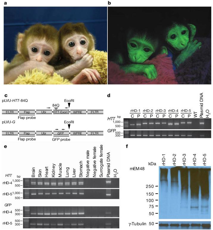
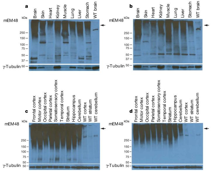
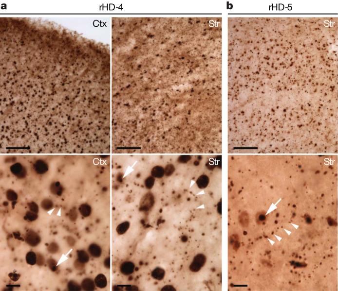
Non-human primates are valuable for modelling human disorders and for developing therapeutic strategies; however, little work has been reported in establishing transgenic non-human primate models of human diseases. Huntington's disease (HD) is an autosomal dominant neurodegenerative disorder characterized by motor impairment, cognitive deterioration and psychiatric disturbances followed by death within 10-15 years of the onset of the symptoms. HD is caused by the expansion of cytosine-adenine-guanine (CAG, translated into glutamine) trinucleotide repeats in the first exon of the human huntingtin (HTT) gene. Mutant HTT with expanded polyglutamine (polyQ) is widely expressed in the brain and peripheral tissues, but causes selective neurodegeneration that is most prominent in the striatum and cortex of the brain. Although rodent models of HD have been developed, these models do not satisfactorily parallel the brain changes and behavioural features observed in HD patients. Because of the close physiological, neurological and genetic similarities between humans and higher primates, monkeys can serve as very useful models for understanding human physiology and diseases. Here we report our progress in developing a transgenic model of HD in a rhesus macaque that expresses polyglutamine-expanded HTT. Hallmark features of HD, including nuclear inclusions and neuropil aggregates, were observed in the brains of the HD transgenic monkeys. Additionally, the transgenic monkeys showed important clinical features of HD, including dystonia and chorea. A transgenic HD monkey model may open the way to understanding the underlying biology of HD better, and to the development of potential therapies. Moreover, our data suggest that it will be feasible to generate valuable non-human primate models of HD and possibly other human genetic diseases.
Figures
Comment in
-
Huntington's disease: genetics lends a hand.Nature. 2008 Jun 12;453(7197):863-4. doi: 10.1038/nature06365. Nature. 2008. PMID: 18488017 No abstract available.
References
-
- Cummings CJ, Zoghbi HY. Trinucleotide repeats: mechanisms and pathophysiology. Annu. Rev. Genomics Hum. Genet. 2000;1:281–328. - PubMed
-
- Myers RH, Marans KS, MacDonald ME. In: Huntington's Disease in Genetic Instabilities and Hereditary Neurological Diseases. Wells RD, Warren ST, editors. Academic; San Diego: 1998. pp. 301–324.
-
- Rubinsztein DC. Lessons from animal models of Hungtington's disease. Trends Genet. 2002;18:202–209. - PubMed
-
- MacDonald ME, et al. HD research collaborative groups. A novel gene containing a trinucleotide repeat that is expanded and unstable on Huntintgton's disease chromosome. Cell. 1993;72:971–983. - PubMed
Publication types
MeSH terms
Substances
Grants and funding
LinkOut - more resources
Full Text Sources
Other Literature Sources
Medical
