. 2008 Jun;10(6):688-97.
doi: 10.1038/ncb1731. Epub 2008 May 18.

STIM1 signalling controls store-operated calcium entry required for development and contractile function in skeletal muscle

Affiliations

STIM1 signalling controls store-operated calcium entry required for development and contractile function in skeletal muscle

Jonathan Stiber et al. Nat Cell Biol. 2008 Jun.

Abstract

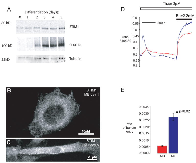
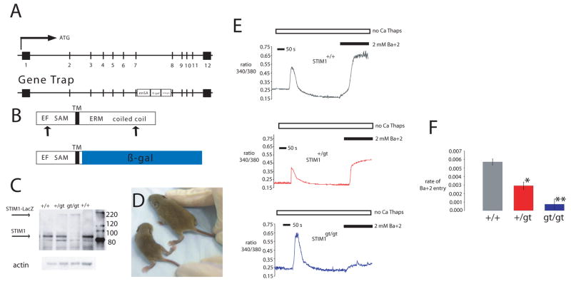
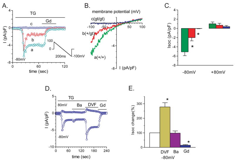
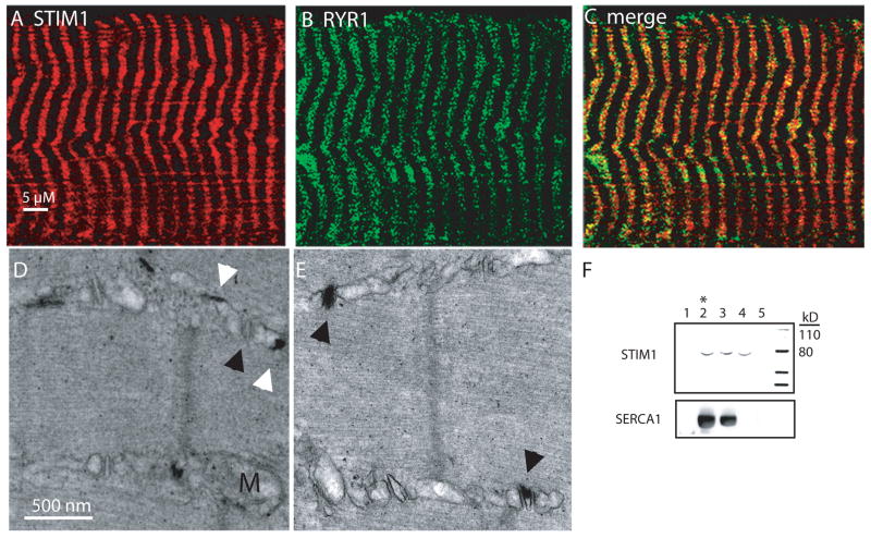
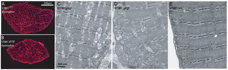
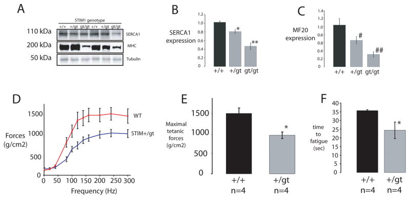
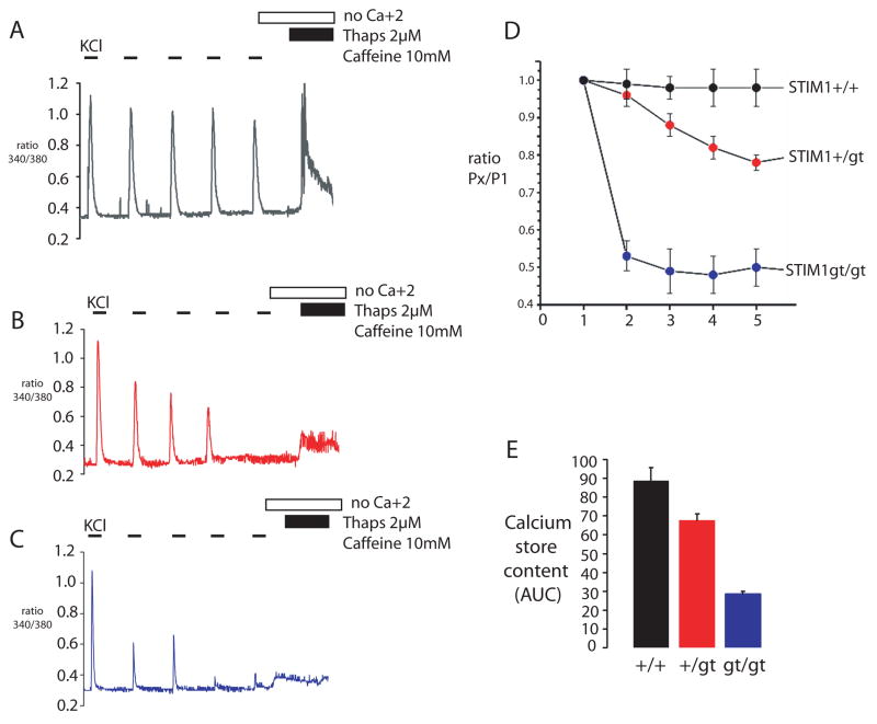
It is now well established that stromal interaction molecule 1 (STIM1) is the calcium sensor of endoplasmic reticulum stores required to activate store-operated calcium entry (SOC) channels at the surface of non-excitable cells. However, little is known about STIM1 in excitable cells, such as striated muscle, where the complement of calcium regulatory molecules is rather disparate from that of non-excitable cells. Here, we show that STIM1 is expressed in both myotubes and adult skeletal muscle. Myotubes lacking functional STIM1 fail to show SOC and fatigue rapidly. Moreover, mice lacking functional STIM1 die perinatally from a skeletal myopathy. In addition, STIM1 haploinsufficiency confers a contractile defect only under conditions where rapid refilling of stores would be needed. These findings provide insight into the role of STIM1 in skeletal muscle and suggest that STIM1 has a universal role as an ER/SR calcium sensor in both excitable and non-excitable cells.


Figures

Figure 1
Figure 1. Muscle differentiation is associated with increased expression of STIM1 and redistribution of STIM1
A) Differentiating C2C12 cells were harvested at the indicated times and protein lysates were separated by SDS-PAGE and immunoblotted for STIM1 and SERCA1 using specific antibodies. Complete scans of these gels are shown in supplemental information (supplemental fig 6). B) STIM1 expression in C2C12 myoblasts (MB, scale bar = 10 μm) and C) in C2C12 cells allowed to differentiate into myotubes (MT, scale bar = 20 μm). STIM1 aggregation and redistribution to the cellular periphery occurs during myogenesis. Arrows represent peripherally localized STIM1. D) Store-operated calcium entry was greater in myotubes than in myoblasts. Fura-2 loaded C2C12 myoblasts and myotubes were placed in zero calcium media, and treated with thapsigargin to induce store depletion and verapamil to inhibit L-type Ca2+ channels. Once cytoplasmic Ca2+ returned to baseline, barium was added to the extracellular medium as a surrogate for Ca2+. Representative average tracings of individual myoblasts and myotubes showed a significant increase in store-operated influx in myotubes. E) The rate of store-operated barium influx, calculated by the first derivative of the 340/380 nm ratio in the first 100 seconds of influx, was 5.57 × 10⁻⁴ ± 4 × 10⁻⁵ arbitrary unit/sec (n = 23) in myoblasts, and 2.72 × 10⁻³ ± 3 × 10⁻⁴ arbitrary unit/sec (n = 6) in myotubes (p<0.001). The data shown represent the mean ± SE.
Figure 2
Figure 2. Gene trap strategy for STIM1
A) Mouse STIM1 gene showing the exon structure in boxes (upper panel). Corresponding STIM1 locus with gene trap vector insertion (lower panel). The gene trap vector carries the engrailed2 intron (en2) and splice acceptor site (SA), β-galactosidase reporter gene and a SV40 polyadenylation site (SV40pA) inserted between exons 7 and 8. B) Both WT and gene-trapped STIM1 protein contain the EF hands and SAM domain of the N-terminus that localizes to the ER lumen, and the membrane spanning region (TM). The cytosolic loop is depicted at the C-terminus only in the WT locus as the gene trap product fuses the first 30 amino acids of STIM1 N-terminus to β-gal. C) Lysates prepared from muscles of WT, +/gt, and gt/gt mice and immunoblotted for STIM1 revealed WT STIM1 and STIM1 fusion protein. Complete scans of these gels are shown in supplemental information (supplemental Fig 6). D) Two week old gt/gt mice appeared smaller and weaker compared to WT littermates. (STIM1gt/gt is seen on the left.) E–F) Store-operated calcium entry in primary myotubes prepared from WT, +/gt, and gt/gt mice. Fura-2 loaded primary myotubes were placed in zero calcium media, and treated with thapsigargin to induce store depletion and verapamil to inhibit L-type Ca2+ channels. Once cytoplasmic Ca2+ returned to baseline, barium was added to the extracellular medium as a surrogate for Ca2+. Representative average tracings of individual myotubes showed a significant decrease in store-operated influx in +/gt and gt/gt myotubes compared with myotubes prepared from WT littermate controls, with minimal influx in gt/gt myotubes.
Figure 3
Figure 3. Store depletion fails to activate SOC current in primary myotubes lacking functional STIM1
SOC currents in response to TG (2μM) were recorded from myotubes prepared from STIM1gt/gt, STIM1+/gt, and STIM1+/+ mice. A) Examples of thapsigargin (TG)-induced Isoc responses in STIM1 gt/gt, +/gt, and +/+ myotubes. The currents were induced by a 200ms voltage ramp protocol (1mV/ms), from 100mV to −100mV, from a holding potential of 0mV (see inset). Sweeps occurred every 2 seconds. Peak Isoc was leak-subtracted and normalized by membrane capacitance. Isoc current density was measured at −80mV. Store-depletion resulted in a large Isoc peak in STIM1 +/+ myotubes (green trace) and a smaller Isoc response in STIM1 +/gt myotubes (red trace), but no significant response in STIM1 gt/gt (blue trace) myotubes. Isoc was inhibited rapidly after the addition of gadolinium (Gd3+, 100μM). B) I/V plots of the Isoc currents after TG perfusion at the times indicated in A) of STIM1 +/+ (a, green trace), STIM1 +/gt (b, red trace), STIM1 gt/gt (c, blue trace). Note the stimulatory effect of TG was absent in STIM1 gt/gt myotubes. C) Group mean values of peak Isoc at −80mV and +80mV in STIM1 +/+ (n=19), STIM1 +/gt (n=8), vs. STIM1 gt/gt (n=8) myotubes; *, P<0.05, STIM1 gt/gt vs. STIM1 +/+, and STIM1 +/gt myotubes. D) An example of TG-induced Isoc response at +80mV and −80mV in solutions containing Ca2+, Ba2+, divalent-free (DVF), and Gd3+ containing solutions respectively. E) Group mean changes of peak Isoc at −80mV in the presence of Ba2+ (maroon bar, n=12), DVF (yellow bar, n=10), and Gd3+ (blue bar, n=9) containing external solutions. Open bars represent control Isoc (100%). *, P<0.05, control versus Ba2+, DVF, or with Gd3+.
Figure 4
Figure 4. STIM1 Localization
A) Immunostaining for STIM1 in skeletal muscle using a STIM1 specific antibody displayed a striated pattern. B) Immunostaining for RYR. C) Merged panel shows partial overlap of STIM1 and RYR. Scale bar (A–C) = 5 μm. D-E) Expression of the STIM1-LacZ fusion protein by electron microscopy. Aggregates of the reaction products of beta-galactosidase were detected in the longitudinal SR (white arrowhead) as well as the junction of the t-tubule and terminal SR (black arrowhead). Scale bar (D–E) = 500 nm. F) Isolated microsomal fractions were obtained using sucrose gradients from rabbit muscle which revealed the absence of STIM1 expression in the fraction corresponding to the contractile proteins and debris (1), and the presence of STIM1 expression in the terminal cisternae (2), longitudinal SR (2 and 3), and t-tubular fractions (4 and 5). * indicates fraction with greatest [H3+] RYR binding. Complete scans of these gels are shown in supplemental information (supplemental Fig 6).
Figure 5
Figure 5. Mice without functional STIM1 display a neonatal skeletal myopathy
A–B) Dystrophin immunostaining of cross sections taken from neonatal muscle of STIM1+/+ and STIM1gt/gt mice. Nuclei were counterstained with DAPI. Scale bar = 200 μm. C–E) Transmission electron microscopy was used to examine muscle ultrastructure from STIM1 gt/gt (C and D) and STIM1+/+ mice (E). TA muscles were taken from 7–10 day old STIM1+/+ and STIM1gt/gt mice. 5000X images were obtained from muscles of two mice. Scale bar = 500 nM.
Figure 6
Figure 6. Muscle gene expression and functional analysis of mutant STIM1 mice
A–C) Muscle protein lysates taken from neonatal mice (STIM1gt/gt, STIM1+/gt versus STIM1+/+) displayed a reduction in SERCA1 (top panel) and Myosin Heavy Chain (middle panel) in mutant STIM1 mice as assessed by immunoblotting with specific antibodies for SERCA1 and MHC (MF20). Quantification using densitometry is provided for studies of three mice for each genotype for SERCA1 (B) and MHC (C). Complete scans of these gels can be found in supplemental information (supplemental Fig 6). D) Contractile force measurements after tetanic stimulation of EDL muscles taken from STIM1+/gt (n=4) and STIM1+/+ mice (n=4). E) Bar graphs represents maximal forces (mean ± SE) following tetanic stimulation for STIM1+/gt and STIM1+/+. F) Bar graph of time to fatigue after repetitive stimulation for muscles taken from STIM1+/gt and STIM1+/+ mice. Time to fatigue was measured using a protocol of one 100Hz stimulation per sec, for a duration of 200ms. Values (mean ± SE) represent the time required for a decay in force generation to 50% maximal force, following stimulation of 4 muscles from each genotype.
Figure 7
Figure 7. STIM1 mediated store refilling is required for fatigue resistance in skeletal myotubes
A–C) Calcium transients were measured from STIM1+/+(A), STIM1+/gt (B) and STIM1gt/gt (C) myotubes by a series of KCl pulses (55mM) in the presence of [Ca+2]o. SR store content was then determined by stimulating myotubes in a zero [Ca+2]o solution with TG (2uM) and caffeine (10mM). D) STIM1+/+ myotubes responded to a series of KCl stimulations with little change in the amplitude of the calcium transient (black trace). STIM1+/gt (red trace) and STIM1gt/gt (blue trace) myotubes responded to the series of KCl-pulses with a decrement in peak amplitude of the calcium transient as measured by the ratio of the amplitude of subsequent KCl pulses to the initial KCl pulse (PX/P1). E) Calcium store content after KCl stimulation in STIM1+/+, STIM1+/gt and STIM1gt/gt myotubes. Data shown represent mean ± SE.

References

    1. Liou J, et al. STIM is a Ca2+ sensor essential for Ca2+-store-depletion-triggered Ca2+ influx. Curr Biol. 2005;15:1235–1241. - PMC - PubMed
    1. Roos J, et al. STIM1, an essential and conserved component of store-operated Ca2+ channel function. The Journal of cell biology. 2005;169:435–445. - PMC - PubMed
    1. Zhang SL, et al. STIM1 is a Ca2+ sensor that activates CRAC channels and migrates from the Ca2+ store to the plasma membrane. Nature. 2005;437:902–905. - PMC - PubMed
    1. Luik RM, Wu MM, Buchanan J, Lewis RS. The elementary unit of store-operated Ca2+ entry: local activation of CRAC channels by STIM1 at ER-plasma membrane junctions. The Journal of cell biology. 2006;174:815–825. - PMC - PubMed
    1. Wu MM, Buchanan J, Luik RM, Lewis RS. Ca2+ store depletion causes STIM1 to accumulate in ER regions closely associated with the plasma membrane. The Journal of cell biology. 2006;174:803–813. - PMC - PubMed

Publication types